Download
1 / 31
340 likes | 894 Views
Anatomy and Electrophysiology of the Heart. By Shomail Sikander. The Heart. The pump of the circulatory system – Contraction pushes blood throughout the body to deliver needed oxygen and nutrients to tissues and remove waste products

E N D
Anatomy andElectrophysiology of the Heart By ShomailSikander
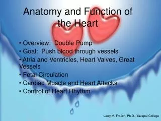
The Heart The pump of the circulatory system – Contraction pushes blood throughout the body to deliver needed oxygen and nutrients to tissues and remove waste products – Depending on body’s requirements, heart rate can either be increased or decreased
The Heart • Shaped like an • inverted blunt cone – Base is the larger, flat part – Apex is the inferior end which tapers to a blunt, rounded point
The Heart • Located between the two lungs in mediastinum behind the sternum
The Heart • Surrounded by pericardial sac (a double-walled closed sac) – fibrous pericardium – serous pericardium
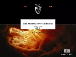
Heart Wall • Made up of three layers – Epicardium (outermost) – Myocardium (middle) – Endocardium (innermost)
Heart Cells • Myocardial cells (working cells) – Contract to propel blood out of heart’s chambers • Electrical conduction system cells – Initiate and carry impulses throughout heart
Myocardial Cells • Cylindrical and branching at their ends – Intercalated disks and gap junctions allow rapid movement of electrical impulses from one cell to another – Desmosomes hold cells together when heart muscle contracts
Internal Heart • Heart consists of four chambers – 2 atria collect blood and deliver to ventricles – 2 ventricles pump blood to pulmonary and systemic circulation • Septum separates heart into two functional units
Heart Valves • Permit blood to flow through heart in only one direction – Mitral and bicuspid • valves (AV valves) located between atria and ventricles – Aortic and pulmonic valves (semilunar valves) located at base of aorta and pulmonary artery
Coronary Arteries • Provide heart with most of its blood supply originate from base of ascending aorta – Immediately above leaflets or cusps of aortic valve
Blood Flow Pulmonary circulation – Pulmonary arteries carry deoxygenated blood to lungs – Pulmonary veins carry oxygenated blood back to heart Systemic circulation • Arteries carry oxygenated blood Veins carry deoxygenated blood
Cardiac Cycle • Diastole – Relaxation and filling of atria and ventricles
Cardiac Cycle Systole – Contraction of atria and ventricles
Cardiac Output • Amount of blood pumped from the heart in one minute – Expressed in LPM
Blood Pressure • The force that blood exerts against walls of arteries as it passes through them • Equals cardiac output times peripheral vascular resistance CO X PVR = BP
Key Properties of MyocardialCells • Automaticity – Can produce electrical activity without outside nerve stimulation • Excitability – Ability to respond to an electrical stimulus • Conductivity – Ability to transmit an electrical stimulus from cell to cell throughout myocardium • Contractility – Ability of myocardial cell to contract when stimulated by an electrical impulse
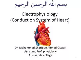
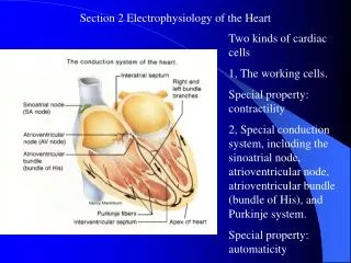
Heart’s Conduction System • Grouping of specialized tissues that carry wave of depolarization throughout heart
Pacemaker Sites • SA node is primary pacemaker site of heart • Other cardiac cells lower in conduction pathway play a back-up role
Polarized State • Inside of myocardial cells more negatively charged in relationship to outside where it is more positively charged
Depolarization • Occurs when positively charged ions move inside cells causing interior to become positively charged – Change in electrical charge over time referred to as cell’s action potential
Repolarization • Follows • depolarization and • occurs when: • – Potassium leaves • cell causing • positive charge to • lower • – Sodium and • calcium are • removed by special • transport systems
Cardiac Cycle • Cardiac cycle begins with RA and LA receiving blood from systemic and pulmonary circulations – Rising pressure within atria forces tricuspid and mitral valves open
Cont…. • Heartbeat initiated by an electrical impulse that arises from SA node • Impulse travels through atria – generates a positive waveform on ECG and contraction of atria
Impulse slows as it passes through AV node from atria to ventricles – Allows atria time to finish filling ventricles
Impulse then rapidly travels through His- Purkinje system – Seen as a flat line following P wave
Depolarization of septum and ventricular walls • generates QRS complex and contraction of ventricles
Repolarization of ventricles is represented on ECG by ST segment and T wave
Thank you & Questions