Download
1 / 123
1.31k likes | 2.12k Views
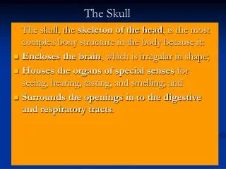
The Skull. Ehab ZAYYAN, MD, PhD. The skull is composed of separate bones united at immobile joints called sutures . Sutural ligaments : between the bones TMJ: the only mobile joint in the skull Skull bones: external and internal tables of compact bone separated by spongy bone called diploë

E N D
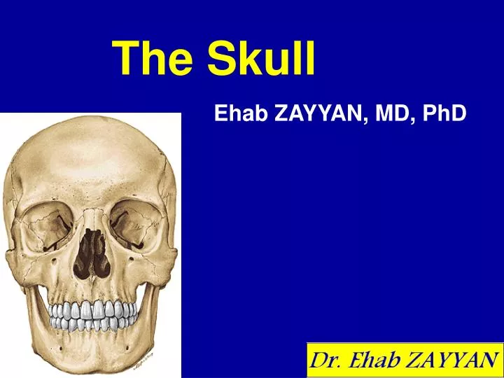
The Skull Ehab ZAYYAN, MD, PhD
The skull is composed of separate bones united at immobile joints called sutures. • Sutural ligaments: between the bones • TMJ: the only mobile joint in the skull • Skull bones: external and internal tables of compact bone separated by spongy bone called diploë • Outer and inner periosteum
Anatomical positionFrankfort horizontal plane The cranium is in the anatomical position when the inferior margin of the orbit and the superior margin of the external acoustic meatus lie in the same horizontal orbitomeatal or Frankfort horizontal plane, a standard craniometric reference
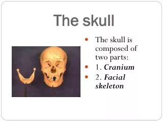
Cranium (Skull) 1. Neurocranium a) Clavaria b) Cranial base 2. Viscerocranium (face bones) Total : 22 bones
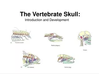
Neurocranium Formed from the mesenchyme of the neural crest Clavariaand skull base (basocranium) • Frontal bone 1 • Parietal bones 2 • Occipital bone 1 • Temporal bones 2 • Sphenoid bone 1 • Ethmoid bone 1
Clavaria • Frontal • Parietal • Occipital • Temporal
Skull base • Occipital • Temporal • Sphenoid • Ethmoid
Frontal • Parietal • Occipital • Temporal • Sphenoid • Ethmoid
Frontal • Ethmoid • Sphenoid • Temporal • Occipital • Parietal
Viscerocranium(Facial skeleton) • Develop from the embryonic mesenchyme of the pharyngeal arches • Consists of the bones surrounding the mouth (upper and lower jaws), nose/nasal cavity, and most of the orbits (eye sockets or orbital cavities)
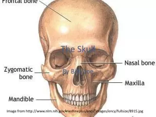
Maxilla • Mandible • Zygoma • Nasalis • Lacrimal
Maxilla • Mandible • Zygoma • Nasalis • Lacrimal
Maxilla • Palatine • Zygoma • Ethmoid and vomer
Mid- sagittal sectionmedial nasal wall • Ethmoid • Vomer • Maxilla • Palatine • Nasalis
Lateral nasal wall • Nasalis, maxilla, ethmoid, inferior concha, palatine
The maxillae contribute the greatest part of the upper facial skeleton, forming the skeleton of the upper jaw, which is fixed to the cranial base. • The mandible forms the skeleton of the lower jaw, which is movable because it articulates with the cranial base at the temporomandibular joints (TMJs).
Several bones of the cranium are pneumatized bones, which contain air spaces (air cells or larger sinuses), presumably to decrease their weight. The total volume of the air spaces in these bones increases with age. Frontal Ethmoid Sphenoid Maxilla Temporal Pneumatized bones of the skull
Pterion (G. wing): Junction of the greater wing of the sphenoid, squamous temporal, frontal, and parietal bones; overlies course of anterior division of middle meningeal artery • Lambda (G. the letter L): Point on calvaria at junction of lambdoid and sagittal sutures • Bregma (G. forepart of head): Point on calvaria at junction of coronal and sagittal sutures • Vertex (L. whirl, whorl): Superior point of neurocranium, in the middle with the cranium oriented in anatomical (orbitomeatal or Frankfort) plane
Asterion (G. asterios, starry): Star shaped; located at junction of three sutures: parietomastoid, occipitomastoid, and lambdoid • Glabella (L. smooth, hairless): Smooth prominence; most marked in males; on the frontal bones superior to root of nose; most anterior projecting part of forehead • Inion (G. back of head): Most prominent point of external occipital protuberance • Nasion (L. nose): Point on cranium where frontonasal and internasal sutures meet
Temporal bone • Squamous • Mastoid • Petrous • Styloid • Tympanic
Neonatal skull At birth the mastoid process and the bony external canal of the tympanic part are absent.
Neonatal temporal bone • Squamous part • Petrous part • Tympanic part
Nasal bones • Anterior nasal aperture
Zygomatic bone • Cheeks prominence • Orbital cavities • Zygomatic arch • Zygomaticofacial & zygomaticotemporal nerve foraminae