Download
1 / 43
580 likes | 1.18k Views
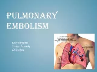
Pulmonary Embolism. Presenter: Dr.Omar Alsherif Moderator: Dr.Hussain Safar. Epidemiology. The related deaths in the United States from venous thromboembolism (VTE) are estimated at 300’000 annually. While in Europe VTE related deaths is estimated at 370’000 per year

E N D
Pulmonary Embolism • Presenter: Dr.Omar Alsherif • Moderator: Dr.Hussain Safar
Epidemiology • The related deaths in the United States from venous thromboembolism (VTE) are estimated at 300’000 annually. While in Europe VTE related deaths is estimated at 370’000 per year • Autopsy studies have shown that approximately 60% of patients who have pulmonary embolism died in the hospital, with the diagnosis having been missed in up to 70% of the cases • DVT occur in 10-13% of patients placed on bed rest for 1 week, 29-33% of all patients in ICU, 20-26% of patients with pulmonary diseases who are given bed rest for > 2 days, 27-33% of patients admitted to CCU after MI, and 48% of patients who under went coronary bypass
Epidemiology • The risk of PE is 20-30% higher in males than in females • Elderly people have higher risks of developing PE than young adults and PE is rare in pediatrics • Pregnant ladies have higher risks of developing PE. The annual incidence is 95.8 per 100’000 women for pregnant ladies and 511.2 per 100’000 women for postpartum ladies. The absolute risk was 199.7 incident per 100’000 women for both • PE accounts for 15% of all postoperative deaths. Lower limb amputations and hip, pelvic, and spinal surgeries are associated with higher risk
Anatomy • Airway • Vascular bed
Anatomy • Airways • Conducting zone, which includes the nose, mouth, pharynx, larynx, trachea, bronchi, bronchioles, and terminal bronchioles. Their function is bring air into and out of the respiratory zone and to warm, humidify, and filter the air • Respiratory zone, which includes the respiratory bronchioles, alveolar ducts, and alveolar sac. Gas exchange occur at that site
Anatomy • Vascular • Pulmonary arteries which is branches into smaller arteries. The smallest arteries divide into arterioles then into capillaries at the respiratory zone
Physiology • Mechanism of inspiration • Inspiration: when the diaphragm and the intercostal muscles contract, the abdominal contents are pushed down and the ribs are lifted upward and outward, increasing the thoracic cavity size and creating -ve pressure. Air will be driven into the lungs • Expiration: it is normally a passive process as air is driven out of the lungs by the reverse pressure gradient between the lungs and atmosphere
Physiology • Gas exchange • According to Fick’s law gas exchange occur due to simple diffusion • In order to maintain the process good capillary perfusion is required
Definition • It is an obstruction of the pulmonary artery usually by blood clot • It is a complication of another disease
Etiology Virchow’s Triad Stasis Thrombosis Hypercoag -ulability Endothelial Injury
Etiology • Venous stasis • Immobilization • Surgery • Obesity • Endothelial injury • Trauma • IV drug abuse • Hypercoagulable states • Pregnancy • Oral contraceptive pills & estrogen replacement therapy • Malignancy • Hereditary
Pathophysiology • There are both respiratory and hemodynamic consequences associated with PE • Respiratory • Increase alveolar dead space ➔ Ventilation-perfusion mismatch ➔ Hypoxemia ➔ Hyperventilation • Hemodynamic • Reduces the cross section of pulmonary vascular bed ➔ Increase pulmonary vascular resistance ➔ Increase Rt ventricle after load ➔ Rt ventricular failure • If left untreated will result in chronic pulmonary hypertension
Clinical Presentation • History • Sudden onset of chest pain • Shortness of breath • Cough, hemoptysis • Palpitations • Cyanosis • Collapse • Sudden death
Clinical Presentation • Examination • Tachypnea • Tachycardia • Fever • Cyanosis • Accentuated second heart sound • S₃ or S₄ gallop • Lower extremity edema • Raised JVP • Tender calf muscle on dorsiflexion of the foot (Homans sign) • Shock
Clinical Scoring Revised Geneva Scoring System
Clinical Scoring • Clinical probability • Score < 4, low risk patient • Score 4-10, intermediate risk patient • Score > 10, high risk patient
Differential Diagnosis • Cardiac • Acute coronary syndrome • Aortic stenosis • Mitral stenosis • CHF • Pulmonary • ARDS • Pneumothorax • Pulmonary edema • COPD & emphysema • Other • Anxiety • Fat embolism
Investigations • Rule In/Out PE • Determine the risk stratification and the management • Clinical evaluation • Cardiac biomarkers • ECG • Echocardiography • CT-Chest
Investigations • Blood tests • CBC • WBC • D-dimer • ABG • S.Troponin I & T • Brain natriuretic peptide (BNP) & NT-pro brain natriuretic peptide (NT-pro BNP) • ECG • Sinus tachycardia • Pulmonary embolism pattern (SI QIII TIII) • prominent S wave in lead I • A Q wave & inverted T wave in lead III • Signs of right ventricular failure • T wave inversion in V₁-V₃ & Qr in V₁ • Peaked P wave in lead II • Right axis deviation • RBBB
Investigations • Radiology • CXR • Atelectasis • Pleural effusion • Prominent central pulmonary artery (knuckle sign) • Cardiomegaly • Pulmonary edema • Wedge shaped triangular opacity (Hampton hump)
Radiology • Echocardiogram • Useful tool for assessing the Rt ventricle function • Assessing the risk stratification • Duplex ultrasound • Helps in demonstrating DVT • -ve scan does not rule out PE as many DVT occur in inaccessible are to U/S • 2/3 of patients with PE caused by DVT has -ve duplex US • Pulmonary angiography • 100% sensitive • 90% specific • Invasive • High dose of radiation • Renal toxicity • High cost • Not always available
Radiology • CT-angiography • The initial imaging modality of choice • Can provide additional information related to alternate diagnosis • Ventilation perfusion scanning • Should only be used when there is contraindication to CT scanning or if CT is not available
Treatment • Primary prevention • Thrombolytic therapy • Surgery • Secondary prevention • Anticoagulant • IVC filter (Greenfield filter)
Treatment • Immediate anticoagulation is mandatory for all patients suspected of having DVT or PE whom hemodynamically stable • Diagnostic investigations should not delay empirical anticoagulant therapy • Anticoagulant must be started with heparin, LMWH, or fondaparinux with warfarin since warfarin requires 5-7 days to achieve therapeutic level
Treatment • Anticoagulant • Heparin; started with IV bolus 5000-10’000 units followed by 1000-1500 units/hour to achieve a target activated partial thromboplastin time APTT 2-3 times the upper limit for the patient • LMWH; enoxaparin (clexane) 1mg/kg SC twice daily • Fondaparinux; 5 mg/day SC for patients < 50 kg, 7.5 mg/day SC for patients 50-100 kg, 10 mg/day SC for patients > 100 kg • Warfarin; starting does 5 mg/day then the dose must be adjusted to achieve INR 2.0-3.0
Treatment • Duration of therapy as recommended by the American College of Chest Physicians • First event of VTE with reversible or time risk factors (trauma, surgery) ⟹ at least three months • First episode of idiopathic VTE ⟹ at least six months • Recurrent idiopathic VTE or continuing risk factor (thrombophilia, spinal cord injury) ⟹ at least one year • Symptomatic isolated calf vein thrombosis ⟹ 6-12 weeks
Treatment • IVC filter implantation indications • Active bleeding • Recurrent venous thrombosis despite intensive anticoagulation
Treatment • Fibrinolysis • The initial management for patients with PE and shock is RESUSCITATION • Successful fibrinolytic therapy is achieved by using 100 mg tissue plasminogen activator (tPA) administered as continuos infusion over two hours • It is contraindicated in intracranial disease, recent surgery or trauma
Treatment • Embolectomy • The American HeartAssociation (AHA) advise that either catheter embolectomy and fragmentation or surgical embolectomy surgical embolectomy is indicated if • Patient has massive PE and contraindication to use thrombolysis • Patient remains unstable after receiving thrombolytic therapy
Prevention • DVT risk stratification level is a method of collecting information from the patient to assess the patient risk of developing DVT
Prevention • The American College of Chest Physicians recommendations • Low risk patient early ambulation • Moderate risk patients • Heparin 5’000 units SC bid or enoxaparin < 40 mg/day, or mechanical prophylaxis • High risk patients • Heparin 5’000 units SC tid or enoxaparin 40 mg/day • Mechanical prophylaxis • Very high risk patients • Adjusted dose heparin, enoxaparin 40 mg bid, or adjusted dose warfarin • Mechanical prophylaxis
Prevention • Mechanical prophylaxis • Thigh high graduated compression stockings • Sequential compression devices • External pneumatic compress • Intermittent pneumatic compression • Venous foot pumps