Download
1 / 13
140 likes | 666 Views
Objectives. Determine some radiopharmaceuticals used.Determine dose ranges Determine the method of administrationList some indications for a Hepatobiliary scan.List contraindications for Hepatobiliary scan.List some patient history needed for Hepatobiliary scan.Trace a radiopharmaceutical from

E N D
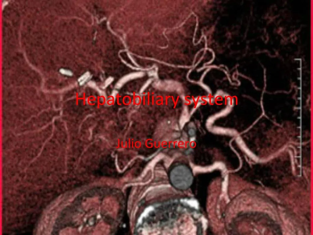
1. Hepatobiliary system Julio Guerrero
2. Objectives Determine some radiopharmaceuticals used.
Determine dose ranges
Determine the method of administration
List some indications for a Hepatobiliary scan.
List contraindications for Hepatobiliary scan.
List some patient history needed for Hepatobiliary scan.
Trace a radiopharmaceutical from injection thru hepatobiliary cycle.
3. Introduction The most common reason for a hepatobiliary scan is to see if a patient suffers from cholecystitis (gallbladder inflammation) or common bile duct obstruction.
Another reason for a scan like this would be for an evaluation of a hepatic transplant.
4. Radiopharmaceuticals the most commonly used radiopharmaceuticals are derivatives of Tc99m Iminodiacetic.
Some radiopharmaceuticals used are Tc99m-IDA (Iminodiacetic ), Tc99m-HIDA
(hepatoimunodiacetic acid), Tc99mDISIDA (diissopropyl imunodiacetic acid)
Typical dose ranges from 5-10mCi
5. Method of Administration IV injection under the camera. Some radiologists want to see how long the heart shadow remains after the injection and perform an immediate review.
6. Indications Evaluation of cholecystitis, or inflammation of cystic or common bile ducts
Evaluation of cholithiasis
Detection of perforation of gallbladder
Evaluation for biliary dyskinesia and congenital anomalies
Evaluation after gallbladder surgery for suspected leakage.
Evaluation of hepatic transplant
7. Contraindications Person has just eaten a meal
No CCK if recently positive for gallstones. Some will administer fatty meal or pulmocare instead as it is not so forceful in gallbladder contraction.
No morphine sulfate if allergic to morphine or has elevated amylase or other pancreatic enzymes indicating pancreatitis.
8. Patient History When was you last solid meal?
Do you have a history or family history of cancer? If so what type and for how long?
Do you have a history of gallbladder or liver disease?
Have you had a recent abdominal pain? If so, where, when, and for how long?
Do you experience nausea or vomiting?
Are you running a fever?
Have you had abnormal blood test results?
Have you had any recent surgery?
Have you had any recent CT, MRI, Ultrasonography or nuclear medicine scans in the abdomen?
Assess for jaundice or signs of alcohol abuse?
9. Procedure Place pt in supine position, camera anterior, liver in upper left quadrant FOV, position liver in middle FOV if heart shadow image,then return liver to LUQ
Image immediate then every 5min up to 30min
Image on dynamic if pt can hold still for 1hr stop dynamic @30-40min if gallbladder seen. Acquire RLAT static.
Gallbladder and bowel visualization: image RAO and RLAT
If no visualization (non-pharmacologic) turn pt to right decubitus position
No visualization (pharmacologic): gall bladder but no bowel, CCK or fatty meal
Bowel but no gallbladder: morphine sulfate,when obtain order from radiologist.
10. Normal results Visualization of liver 5-15sec after injection, hepatic and common bile duct and gallbladder 5-20min up to 1hr.
Intestinal activity visualizing and moving within 10-60min
A flow study will show liver immediately but dimely from activity entering thru hepatic artery, then brightly thru portal vein flow.
Liver diminishes in activity as gall bladder visualizes in its bed and grows brighter and bowel activity visualizes and moves with time, bile ducts in liver appears as lines leading to bowel.
Post-gallbladder surgery, there should be no activity pooling around liver in abdomen.
11. Abnormal results Nonvisualization of gallbladder within 1hr with visualization of common bile duct and bowel (indicates cystic duct obstruction,chlecytitis)
Non-visualization of bowel 1hr with good hepatic uptake, visualization of gallbladder and common bile duct (possible sphincter of oddi dysfunction or obstruction)
Non-visualization of gallbladder or bowel within 1hr with good hepatic uptake but no draining (complete or near complete obstruction of hepatic duct dysfunction)
12. Tracing the Radiopharmaceutical 1.Rt and LT hepatic duct
2.Common hepatic duct
3.Cystic duct
4.Common bile duct
5.duodenum
13. Normal scan