Download
1 / 15
150 likes | 400 Views
Intrahepatic Shunts. James Montgomery, DVM March 30, 2009 Acc # 111557 & 111556. Acc#: 111557. Madeline 3 month old female basset hound Cystoliths Elevated liver enzymes Elevated bile acids Referred for evaluation of shunt. Acc#: 111557. Acc#: 111556. Acc#: 111557. Portal Vein.

E N D
Intrahepatic Shunts James Montgomery, DVM March 30, 2009 Acc # 111557 & 111556
Acc#: 111557 • Madeline • 3 month old female basset hound • Cystoliths • Elevated liver enzymes • Elevated bile acids • Referred for evaluation of shunt
Portal Vein • Formed within the mesentery dorsal to the right limb of the pancreas • Confluence of cranial and caudal mesenteric portal branches • Receives blood from the GI tract and spleen • Mesenteric veins, gastroduodenal, splenic, and gastric veins • As approaches liver, sweeps from right to left and divides into right and left branches • Main right branch – right lateral and caudate process of caudate lobe • Main left branch – all other lobes
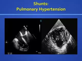
Intrahepatic Shunts • Almost all intrahepatic shunts occur in large dogs • Connect portal vein branches after their divergence from the portal vein to the hepatic veins or abdominal vena cava, bypassing the hepatic sinusoids • Many intrahepatic shunts result from failure of the ductus venosus to close in infancy • 65% are anatomically considered PDV • Divided into right, left, and central divisional shunts
Fetal Circulation Dyce, Sack & Wensing, Textbook of Veterinary Anatomy, 2nd ed, p. 249.
Intrahepatic Shunts • Right divisional shunts • Pass through either the caudate process of the caudate lobe or the right lateral lobe of the liver before entering the vena cava • Central divisional shunts • Pass through either the right medial or quadrate lobes before entering the vena cava • Left divisional shunts • Pass through either the papillary process of the caudate lobe, the left lateral lobe or the medial lobe before entering the vena cava
Madeline 1 2 3
Madeline R Miller’s Anatomy of the Dog, 3rd ed, p. 453. Acc#: 111557
Treatment • Often requires more than one surgery • Surgical techniques described • Posthepatic left hepatic vein attenuation – no clinical effect • Posthepatic direct shunt ligation • Intrahepatic direct shunt dissection and ligation with ultrasound guidance • Intravascular placement of embolisation coils • Intraluminal closure of the shunt via the thoracic vena cava • Transportal closure of the shunt via portal venotomy • Direct attenuation of the PDV before it enters the venous ampulla is technically less demanding
Treatment • The significant blood flow through large intrahepatic shuts cannot be suddenly obstructed • Life threatening portal hypertension would develop • Causes of intraoperative death • Intraoperative shock (hemorrhagic and septic), cardiac arrest, and portal hyper- and hypotension • Postoperative survival rates: 75-89% • Peritonitis, portal vein thrombosis, and portal hypertension are most common postoperative causes of death
Prognosis • Predictors for postoperative complications • Low body weight (<10 kg) and hypoproteinemia or hypoalbuminemia • Shunt location had no effect on short or long-term outcome • Partially attenuated shunt survival rates • 1 year – 60% • 2 year – 55% • After 4 months, euthanasia due to failure to show clinical improvement was most common cause of death
References • Mathews KG, Bunch SK. Vascular Liver Diseases. In Ettinger SJ, Feldman EC, eds. Textbook of Veterinary Internal Medicine, 6th ed (St. Louis, MO: Elsevier, 2005) pp. 1453-64. • White RN, Burton CA. Anatomy of the patent ductus venosus in the dog. Veterinary Record (2000) 146, 425-9. • Whiting PG, Peterson SL. Portosystemic shunts. In Slatter D, ed. Textbook of Small Animal Surgery, 2nd ed (Philadelphia, PA: WB Saunders Co., 1993) pp. 660-74.