Download
1 / 50
520 likes | 595 Views
Organogenesis Part 2. V. Lateral Plate Mesoderm VI. Endoderm VII. Development of the Tetrapod Limb VIII. Sex Determination. V. Lateral Plate Mesoderm. paraxial mesoderm. chordamesoderm. intermediate mesoderm. lateral plate mesoderm. Lateral Plate Mesoderm. Terminology:

E N D
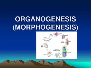
Organogenesis Part 2 V. Lateral Plate Mesoderm VI. Endoderm VII. Development of the Tetrapod Limb VIII. Sex Determination
V. Lateral Plate Mesoderm paraxial mesoderm chordamesoderm intermediate mesoderm lateral plate mesoderm
Lateral Plate Mesoderm Terminology: - Somatopleure: somatic mesoderm plus ectoderm - Splanchnopleure: splanchnic mesoderm plus endoderm - Coelom: body cavity forms between them
Lateral Plate Mesoderm • The Coelom: • eventually left and right cavities fuse into one • runs from neck to anus in vertebrates • portioned off by folds of somatic mesoderm • pleural cavity: surrounds the thorax and lungs • pericardial cavity: surrounds the heart • peritoneal cavity: surrounds the abdominal organs
Figure 12.1 Mesodermal development in frog and chick embryos (Part 3)
Heart Development • The heart is the first organ to function in the embryo and the circulatory system is the first functional system. • heartarteriescapillariesveinsheart • Before the embryo can get very big it must switch from nutrient diffusion to active nutrient transport
Heart Development Anatomical Stages: • Tube Formation • Looping • Chamber Formation outflow inflow human timeline
Heart Development: Tube Formation presumptive heart cells are specified but not determined in the epiblast outflow forming cells (red) migrate in first, inflow second migrate through together near node “the heart field”
Heart Development: Tube Formation The cardiogenic mesoderm migrates out of the mesodermal layer towards the endoderm to form endocardial tubes on either side. At the same time the endoderm is folding inward
Heart Development: Tube Formation The endoderm continues folding inward until it forms its own tube, which drags the two endocardial primordia close to each other. The endocardial tubes are surrounded by myocardial progenitors When the endocardial tubes get close enough, they fuse together
Heart Development: Tube Formation If you mess with endoderm migration or signaling, you end up with two hearts
Heart Development: Tube Formation • Heart Tube Cell Biology • Splanchnic mesoderm cells express cadherins and form an epithelial sheet for their inward migration - MET • The presumptive endocardial cells undergo EMT to migrate away from the sheet and another MET to form tubes • The cells in the original mesodermal sheet form the myocardium • The myocardial epithelium fuses first and the two endocardial tubes exist together inside for a while before fusing • Both the rostral end (outflow) and caudal end (inflow) remain as unfused double tubes • The heart beat starts spontaneously as myocardial cells express the sodium-calcium pump - before fusion is even complete
Heart Development: Looping and Chamber Formation left-right asymmetry is due to Nodal and Pitx2 anterior posterior right left Looping requires: cytoskeletal rearrangement extracellular matrix remodeling asymmetric cell division
Heart Development: Looping and Chamber Formation valve formation heart valves keep the blood from flowing back into the chamber it was just ejected from The septa separate the two atria and the two ventricles septation
Heart Development: Looping and Chamber Formation The truncus arteriosis, or outflow tract, also becomes septated allowing one great artery to flow from right ventricle to lungs and the other from left ventricle to the body.
Heart Development: Looping and Chamber Formation • The tricuspid valve is between the right atrium and right ventricle. • The pulmonary or pulmonic valve is between the right ventricle and the pulmonary artery. • The mitral valve is between the left atrium and left ventricle. • The aortic valve is between the left ventricle and the aorta.
Heart Development: Looping and Chamber Formation Steps: 1. Endocardial cushions form and fuse 2. Septa grow towards cushion 3. Valves form from myocardium In utero, the foramen ovale allows right left shunting of blood
Embryonic circulatory systems All of the blood must circulate outside of the embryo for oxygenation
Blood Vessel Development • The vessels form independently of the heart • They form for embryonic needs as much as adult • Must get nutrition before there is a GI tract • Must circulate oxygen before there are lungs • Must excrete waste before there are kidneys • They do these through links to extraembryonic membranes
Blood Vessel Development • The vessels are constrained by evolution • Mammals still extend vessels to empty yolk sac • Birds and mammals also build six aortic arches as if we had gills, eventually settling on a single arch • The vessels adapt to the laws of fluid dynamics • Large vessels move fluid with low resistance • Diffusion requires small volumes and slow flow • Highly organized size variance controls volume • And superbranching smaller vessels control speed
Blood Vessel Development Vasculogenesis is the de novo differentiation of mesoderm into endothelium It is followed by the endothelium recruiting smooth muscle cell coat
Blood Vessel Development Starts in the extraembryonic mesoderm as well as in the large embryonic blood vessels
Blood Vessel Development Angiogenesis is the growth and remodeling of the 1st vessels in response to blood flow and tissue-derived recruitment signals
Blood Vessel Development Secondary Vasculogenesis 1. PEO forms from splanchnic mesoderm overlying the liver 2. PEO contacts the ventricle and migrates as epicardium 3. Subset of epicardial cells delaminate towards myocardium 4. These undergo MET to form coronary endothelium 5. Coronary arteries then plug into the aorta where nerves are
Blood Vessel Development It is a common phenomenon for arteries and nerves to form together Less so for veins....
Blood Vessel Development • Lymphatic drainage forms from jugular vein • Sprouts as lymphatic sacs by angiogenesis • Continues to form secondary drainage system • Major conduit for immune cells
Where do the hematopoietic stem cells of the adult bone marrow come from? Splanchnic mesoderm of aorta-gonad-mesonephros (AGM) region in embryo Hemogenic endothelium from sclerotome Hemogenic endothelium from many sites
Development of the Endoderm • The Digestive Tube • Anterior endoderm forms anterior intestinal portal • Posterior endoderm forms posterior intestinal portal • Midgut goes through expansion and contraction to yolk • Each end has ectodermal cap, then forms an entrance • The Derivatives • 4 pharyngeal pouches form head and neck structures • Floor between 4th pair buds out to form respiratory tube • Gut tube forms esophagus, stomach, SI, LI, rectum • Gut tube buds out to form liver, gall bladder, pancreas
Development of the Endoderm Human Timeline
Development of the Endoderm The cranial neural crest cells migrate through this endoderm and contribute component structures around them
Development of the Endoderm Localized Wnt/B-Catenin and retinoic acid cause budding
Development of the Endoderm Normal-time birth is signaled from the lungs
Development of the Endoderm Anterior-Posterior specification of the gastrointestinal tract
Development of the Endoderm Reciprocal Induction Simultaneous Anterior-Posterior specification of both endoderm and mesoderm
Development of the Endoderm Mesoderm also induces liver bud
The Extraembryonic Membranes • Adaptation for development on dry land • As the body starts to develop epithelium expands to isolate embryo within them • Four sets of extraembryonic membranes • Somatopleure forms amnion and chorion • Splanchnopleure forms yolk sac and allantois
The Extraembryonic Membranes • Somatopleure forms amnion and chorion • Splanchnopleure forms yolk sac and allantois
The Extraembryonic Membranes The amnion folds up to cover the embryo and keep it from drying out The cells of the amnion secrete water
The Extraembryonic Membranes The chorion surrounds the entire embryo and controls gas exchange In birds and reptiles it lines shell In mammals it forms the placenta
The Extraembryonic Membranes The yolk sac expands to surround yolk (even if you don’t have any)
The Extraembryonic Membranes The allantoic membrane creates a space for waste storage Bird and reptile eggs gotta’ have it We don’t use it for waste but it contributes to our umbilical cord