Download
1 / 25
280 likes | 437 Views
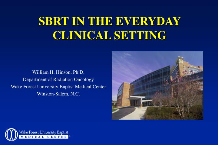
SBRT IN THE EVERYDAY CLINICAL SETTING. William H. Hinson, Ph.D. Department of Radiation Oncology Wake Forest University Baptist Medical Center Winston-Salem, N.C. SBRT Team at WFUBMC. Physician Volker Stieber William Blackstock Kevin McMullen Physics William H. Hinson William T. Kearns

E N D
SBRT IN THE EVERYDAY CLINICAL SETTING William H. Hinson, Ph.D. Department of Radiation Oncology Wake Forest University Baptist Medical Center Winston-Salem, N.C.
SBRT Team at WFUBMC Physician • Volker Stieber • William Blackstock • Kevin McMullen Physics • William H. Hinson • William T. Kearns Whole cast of support staff
Essentials for SBRT • 3-D Imaging device • Treatment planning system • Treatment delivery system • Precise targeting (stereotactic coordinate system) • Patient positioning verification
Specialized Devices for SBRT • Novalis • Cyberknife • Accelerator-based IGRT (Trilogy, Synergy)
Essentials for SBRT • 3-D Imaging device • Treatment planning system • Treatment delivery system • Precise targeting (stereotactic coordinate system) • Patient positioning verification
WFUBMC SBRT3-D Imaging Device • Phillips PQ 5000 CT simulator • Single slice acquisition • External lasers for isocenter placement • AcQSim software
WFUBMC SBRT“New” 3-D Imaging Device • GE Discovery ST CT/PET Simulator • 8 slice, helical scan, LightSpeed (Ultra) CT scanner • External lasers for isocenter placement • Advantage Simulation Workstation software
WFUBMC SBRTTreatment Planning System • Phillips-ADAC 3D RTP system • Accurate small field dosimetry • Standard plan consists of 9 coplanar beams with negative margins
WFUBMC SBRT • Varian 2100 SCX accelerator • 6MV photon beam • 120 leaf MLC • Portal Vision
WFUBMC SBRTStereotactic Patient Positioning • Elekta Stereotactic bodyframe • Cartesian coordinate fiducials • Diaphragm breathing control • Patient must pass “YMCA” test • “Treat the box” approach
Stereotactic Patient Positioning • Precise targeting requires a stereotactic coordinate system or image-based targeting • Patient positioning verification is a MUST.
WFUBMC SBRTPatient Repositioning Verification • Elekta bodyframe lasers (2 chest points plus leg marker • Serial CT image sets on day of simulation • “GTV” is defined from the fusion of 2 image sets (i.e. “4-D GTV”)
WFUBMC SBRTPatient Repositioning Verification • On day of treatment - SSD checks and orthogonal isocenter portal images • Physician approval of isocenter images required before treatment proceeds. • Total time on linac is about 1 to 1.5 hours
WFUBMC SBRTOrthogonal Portal Images Fraction #1 Fraction #2 Fraction #3
WFUBMC SBRTPatient Positioning Verification • Neuro-surgical Stealth Station computer system • Stereo IR camera • Fiducials placed on SBF and patient • System detects shifts in patient, relative to the SBF
WFUBMC SBRTPatient Positioning Verification *Shifts mainly in axial direction on 3mm thick slices.
WFUBMC SBRT • Through March, 2005, 70 SBRT patients have been treated using the Elekta Bodyframe. • Of these 70 patients, 30 patients have been treated in a single fraction on our Stereotactic Body Radiosurgery protocol. • Others have received between 2 and 4 fractions.
WFUBMC SBRS PROTOCOL CCCWFU 99502: “A Phase I/II Dose-Escalation/Efficacy Study of Palliative Extracranial Radiosurgery using the Elekta Stereotactic Body Frame System.” Eligibility criteria: • The presence of a well-circumscribed tumor (primary or metastatic) on contrast-enhanced CT scan with a maximum diameter of 6 cm. • Age 18 years with a life expectancy 3 months. • No chemotherapy or immunotherapy were allowed at least 3 weeks prior to or planned for 4 weeks after treatment.
WFUBMC SBRS PROTOCOL • Patients were stratified to dose levels by tumor volume • Phase I Endpoint: Acute toxicity using NCI Common Toxicity Criteria 3.0 • Phase II Endpoint: Local control by CT RECIST Criteria (Response Evaluation Criteria In Solid Tumors)
WFUBMC SBRS PROTOCOL • 28 patients have been enrolled on study. 12 (43 %) of these patients have had PET imaging for a minimum of 3 months. We report the results of these patients. • For the PET group, median follow-up so far is 377 days (171-739 days). Median survival has not yet been reached. 3 deaths have occurred. For these 3 patients, median survival from time of treatment was 250 days.
WFUBMC SBRS PROTOCOL • Local control at 3 months was 100 %. • The median change in tumor diameter at 3 months was -14 % (-59% to 0 %) for a median RECIST of SD. • The median corrected SUV change at 3 months was -57 % (-13% to + 11%).
WFUBMC SBRS PROTOCOL • 8 patients have had no progressive disease (PD). • For 7/8, the SUV decreased and had not increased by the last follow-up. • 4 patients have had PD by RECIST. • 3/4 had a corresponding rise in correct SUV, which preceded the RECIST progression by a median of 71 days (71-75 days). • The median time to RECIST failure for these patients was 238 days (175-319 days). • Overall, the correlation between RECIST control and PET control was 0.71.
WFUBMC SBRS PROTOCOL • In the acute phase (< 3 months), there appears to be no significant inflammatory response by PET. • In the late phase, (> 3 months) PET progression appears to precede RECIST PD by 2.4 months. This may have implications for the timing of subsequent therapy (e.g. chemotherapy).
POSSIBLE IMPROVEMENTS • Respiratory gating • On-board KV imaging • Cone beam imaging