Download
1 / 118
1.23k likes | 1.38k Views
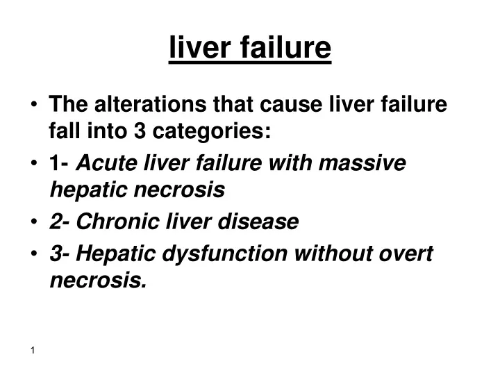
liver failure. The alterations that cause liver failure fall into 3 categories: 1- Acute liver failure with massive hepatic necrosis 2- Chronic liver disease 3- Hepatic dysfunction without overt necrosis. 1-Acute liver failure.

E N D
liver failure • The alterations that cause liver failure fall into 3 categories: • 1- Acute liver failure with massive hepatic necrosis • 2- Chronic liver disease • 3- Hepatic dysfunction without overt necrosis.
1-Acute liver failure. • This is most often caused by drugsor fulminant viral hepatitis. • Acute liver failure denotes clinical hepatic insufficiency that progresses from onset of symptoms to hepatic encephalopathy within 2 to 3 weeks. • A course extending as long as 3 months is called subacute failure.
The histologic correlate of acute liver failure is massive hepatic necrosis. • It is an uncommon but life-threatening condition that often requires liver transplantation.
2-Chronic liver disease • This is the most common route to hepatic failure and is the end point of relentless chronic liver damage ending in cirrhosis.
Hepatic dysfunction without overt necrosis. • Hepatocytes may be viable but unable to perform normal metabolic function: • 1- acute fatty liver of pregnancy (which can lead to acute liver failure a few days after onset) • 2- tetracycline toxicity • 3- Reye syndrome
Clinical features 1-Jaundice 2-Hypoalbuminemia →edema 3-Hyperammonemia 4-Fetor hepaticus (musty or sweet & sour) 5-Palmar erythema hyperestrogenemia 6-Spider angiomas 7-Hypogonadism & gynecomastia
Complications: 1-Multiple organ failure e.g lung 2-Coagulopathy → bleeding def. factors II, VII, IX, X 3-Hepatic encephalopathy 4-Hepatorenal Syndrome
Alcoholic liver disease -Alcohol is most widely abused agent -Excessive ethanol consumption causes more than 60% of chronic liver disease in most Western countries and accounts for 40% to 50% of deaths due to cirrhosis. -It is the 5th leading cause of death in USA due to : 1.Accident 2.Cirrhosis
Pathogenesis • Short-term ingestion of as much as 80 gm of ethanol/d (8 beers or 7 ounces of 80-proof liquor) generally produces mild, reversible hepatic changes. • Chronic intake of 50 to 60 gm/day is considered a borderline risk for severe injury. • women seem to be more susceptible to hepatic injury than are men because of low gastric metabolism of ethanol and differences in body composition.
-80 – 100 mg/dl is the legal definition for driving under the influence of alcohol -44 ml of ethanol is required to produce this level in 70kg person -In occasional drinkers, bl. Level of 200 mg/dl produces coma & death & respiratory failure at 300-400 mg/dl
Habitual drinkers can tolerate levels up to 700 mg/dl without clinical effect due to metabolic tolerance explained by 5-10X induction of cytochrome P-450 system that includes enzyme CYP2E1 which increases the metabolism of ethanol as well as other drugs as cocaine & acetominophen
Forms of alcoholic liver disease 1-Hepatic steatosis (90-100% of drinkers) 2-Alcoholic hepatitis ( 1- 35% of drinkers) 3-Cirrhosis ( 14% of drinkers) • Steatosis & hepatitis may develop independently
Hepatic steatosis -Can occur following even moderate intake of alcohol in form of microvesicular steatosis • initially centrilobular but in severe cases it may involve the entire lobule . -Chronic intake → diffuse steatosis -Liver is large ( 4 – 6 kg) soft yellow & greasy -Continued intake →fibrosis -Fatty change is reversible with complete absention from further intake of alcohol
Alcoholic hepatitis Characteristic findings : 1-Hepatocyte swelling & necrosis -Accumulation of fat & water & proteins -Cholestasis -Hemosiderin deposition in hepatocytocytes & kupffer cells 2-Mallory-hayline bodies -eosinoplilic cytoplasmic inclusions in degenerating hepatocytes formed of cytokeratin infermediate filaments & other proteins
-Mallory-hayline inclusions are characteristic but not pathognomonic of alcoholic liver disease, they are also seen in : 1-Primary biliary cirrhosis 2-Wilson disease 3-Chronic cholestatic syndromes 4-Hepatocellular carcinoma
3-Neutrophilic reaction 4-Fibrosis -Sinusoidal & perivenular fibrosis -Periportal fibrosis 5-Cholestasis 6-Mild deposition of hemosiderin in hepatocytes & kupffer cells
Alcoholic cirrhosis -Usually it develops slowly -Initially the liver is enlarged yellow but over years it becomes brown shrunken non-fatty organ s.t < l kg in wt. -Micronodular → mixed micro & macronodular -Laennec cirrhosis = scar tissue -Bile stasis -Mallory bodies are only rarely evident at this stage -Irreversible -It can develop rapidly in the presence of alcoholic hepatitis (within 1-2 yrs).
Ethanol metabolism Ethanol → acetaldehyde CH3 CH2OH CH3 C=O H ↑ -Alcohol dehydrogenase (stomach + liver) -Cytochrome P-450 -Catalase ( liver) -
Acetaldehyde → Acetic acid ↑ Aldehyde dehydrogenase
After absorption ethanol is distributed as Acetic acid in all tissues & fluid in direct proportion to blood level • Women have lower levels of gastric alcohol dehydrogenase activity than men & they may develop higher blood Levels than men after drinking the same quantity of ethanol.
- Less than 10% of absorbed ethanol is excreted unchanged in urine , sweat & breathe -There is genetic polymorphism in aldehyde dehydrogenase that affect ethanol metabolism e.g 50% of chinese , vietnamase & Japanese have lowered enzyme activity due to point mutation of the enzyme. → accumulation of acetaldehyde → facial flushing, tachycardia & hyperventilation. • -
Clinical features -Hepatic steatosis ( reversible ) ↑ liver ↑ liver enz. Severe hepatic dysfunction is unusual -Alcoholic hepatitis • 15-20 yr. of excessive drinking • Non-specific symptoms, malaise, anorexia, wt. loss • Hepatosplenomegaly • ↑ LFT Each bout of hepatitis →10-20% risk of death → cirrhosis in 1/3 in few yrs. -Cirrhosis Portal hypertension
Causes of death in alcoholic liver disease 1-hepatic failure 2-Massive GI bleeding 3-Infections 4-Hepatorenal syndrome 5-HCC in 3-6% of cases
Cirrhosis • It is a diffuse process characterized by fibrosis & the conversion of liver parenchyma into nodules
Main characteristics 1.Bridging fibrous septae 2.Parenchymal nodules encircled by fibrotic bands 3.Diffuse architecture disruption
Types : Micronodules < 3mm in diameter Macronodules > 3 mm in diameter
Causes of cirrhosis 1.Chronic alcoholism 2.Chronic viral infection HBV & HCV 3.Biliary disease 4.Hemochromatosis 5.Autoimmune hepatitis 6.Wilson disease 7.α-1- antitrypsin deficiency
8. Rare causes Galactosemia Tyrosinosis Glycogen storage disease III &IV Lipid storage disease Hereditary fructose intolerance Drug induced e.g methyldopa 9. Cryptogenic cirrhosis 10%
Pathogenesis of cirrhosis -The mechanism of cirrhosis involves: 1-Hepatocellular death 2-Regeneration 3-Progressive fibrosis 4-Vascular changes
The development of cirrhosis requires that cell death occur over long periods of time and be accompanied by fibrosis. • Fibrosis progresses to scar formation when the injury involves not only the parenchyma but also the supporting connective tissue.
-In normal liver the ECM collagen (types I, III,V & XI) is present only in : Liver capsule Portal tracts Around central vein
-delicate framework of type IV collagen & other proteins lies in space of Disse -In cirrhosis types I & III collagen & others are deposited in the space of Disse
Vascular changes consisting of the loss of sinusoidal endothelial cell fenestrations and the development of portal vein-hepatic vein and hepatic artery-portal vein vascular shunts contribute to defects in liver function.
Collagen deposition converts sinusoids with fenestrated endothelial channels that allow free exchange of solutes between plasma and hepatocytes to higher pressure fast-flowing vascular channels without such solute exchange.
The movement of proteins (e.g., albumin, clotting factors, lipoproteins between hepatocytes and the plasma is markedly impaired. • These functional changes are aggravated by the loss of microvilli from the hepatocyte surface which diminishes the transport capacity of the cell.
- The major source of collagen in cirrhosis is the perisinusoidal stellate cells (Ito cells) which lie in space of Disse - Perisinusoidal stellate cells act normally as storage cells for vit A & fat
Activated stellate cells produce growth factors, cytokines, and chemokines that cause their further proliferation and collagen synthesis. • TGF-β is the main fibrogenic agent for stellate cells.
Fibrosis is a dynamic process that involves the synthesis and deposition of ECM components activation of inhibitors of metalloproteinases
-The stimuli for the activation of stellate cells & production of collagen are : 1-reactive oxygen species 2-Growth factors 3-cytokines TNF, IL-I, lymphotoxins
-Clinical features of cirrhosis : -Silent -Anorexia, wt loss, weakness -Complications : 1-Progressive hepatic failure 2-Portal hypertension 3-Hepatocellular carcinoma
Portal hypertension • ↑ resistance to portal blood flow at the level of sinusoids & compression of central veins by perivenular fibrosis & parenchymal nodules • Arterial – portal anastomosis develops in the fibrous bands →increase the blood pressure in portal venous system
Causes of portal hypertension I.Prehepatic 1-Portal vein thrombosis 2-Massive splenomegaly II. Post hepatic 1-Severe Rt.- sided heart failure 2-Constrictive pericarditis 3-Hepatic vein out flow obstruction III. Hepatic 1-Cirrhosis 2-Schistosomiasis 3-Massive fatty change 4-Diffuse granulomatosis as sarcoidosis, TB 5-Disease of portal microcirculation as nodular regenerative hyperplasia
Clinical consequence of portal hypertension 1-Ascitis 2-Portosystemic shunts 3-Hepatic encephalopathy 4-Splenomegaly
Ascitis -Collection of excess fluid in peritoneal cavity -It becomes clinically detectable when at least 500 ml have accumulated -Features 1-Serous fluid 2-Contains as much as 3g/ml of protein (albumin) 3-It has the same concentration as blood of glucose, Na+, & K+ 4-Mesothelial cells & lymphocytes 5-Neutrophils = infection 6-RBCs = DISSEMINATED CANCR
-Pathogenesis 1-Sinusoidal ↑ Bp 2-Hypoalbuminemia 3-Leakage of hepatic lymph into the peritoneal cavity N- thoracic duct lymph flow is 800-1000 ml/d in cirrhosis it may approach 20L /day 4-Renal retention of Na+ & water due to 2ry hyperaldosteronism
Portosystemic shunt -Because of ↑portal venous pressure bypasses develop wherever the systemic & portal circulation share capillary beds -Sites: 1-Around & within the rectum (Hemorrhoids) 2-Gastroesophageal junction (varicies ) 3-Retroperitoneum 4-Falciform ligament of the liver (periumbilical & abdominal wall collaterals ) → caput medusae
- Gastroesophageal varicies appear in 65% of pts. with advanced cirrhosis & cause death in 50% of then due to UGI bleeding
Splenomegaly -Usu. 500-1000 gms (N <300gms) -Not necessarily correlated with other features of portal ↑Bp -May result in hypersplenism
Hepatic Encephalopthy -It is a complication of acute & chronic hepatic failure -Disturbance in brain function ranging from behavioural changes to marked confusion & sutpor to deep coma & death -The changes may progress over hrs. or days
-Neurological signs: Rigidity Hyper-reflexia Non – specific EEG Seizures Asterixis ( non-rhythmic rapid extension flexision movements of head & extremities) . -Brain shows edema & astrocytic reaction