Download
1 / 47
470 likes | 485 Views
Acute Renal Failure (ARF) is a complex syndrome characterized by a sudden decline in renal function. Learn about the definition, classifications (prerenal, renal, postrenal), and management strategies. Understand the significance of biomarkers, epidemiology, and specific syndromes such as Acute Tubular Necrosis (ATN). Find out about the impact of medications like ACE inhibitors and NSAIDs on ARF and explore atypical causes like Abdominal Compartment Syndrome. Discover how to differentiate and address different types of ARF for optimal patient outcomes.

E N D
Acute Renal Failure Syed Rizwan, MD
Acute Renal Failure • Comprises a family of syndromes • Abrupt decrease in GFR(over hours to days)
MANIFFESTATIONS of ARF • Increase in BUN • Increase in creatinine • Oligouria(< 400 –500 cc)
DEFINITION • No consensus • Multiple • Relative rise in Serum Creatinine • > 0.5mg/dl if baseline creatinine is normal • > 1 mg/dl if baseline serum creatinine is high
Creatinine and GFR • Creatinine produced in muscles • Creatinine excretion depends on, • Glomerular filtration • Proximal tubular excretion • Change in Serum Creatinine with no change in GFR • Muscle wasting or amputation lowers creatinine • Medications(Trimethoprim, Cimetidine) increase creatinine by deceasing tubular excretion
Blood Urea and GFR • Increase BUN with no change in GFR • GI Bleed • Hyper catabolic states • Protein loading • Glucocorticoids • Tetracycline • Decrease BUN with no change in GFR • Protein Malnutrition • Severe Liver disease
ARF and Biomarker • Lack of sensitivity of BUN and creatinine • Need for Biomarkers • Kidney Injury Molecules-1(KIM-1) increased in Patients with Acute Tubular Necrosis • None available for cliniical utility yet
Epidemiology of ARF • Incidence, etiology and outcome varied depending on Population studied and Definition used • Mostly in-Patient than out –Patient • 5-7% of hospital admissions • Mortality varies between 20%-85% depending on cause
ARF Classification • Prerenal • Renal • Postrenal
Prerenal ARF • Hemodynamically mediated reduction in GFR in absence on Renal Parenchymal injury. • ARF resolves if hemodynamic insult is reversed • If hemodynamic insult is sustained, can result in overt renal injury
Renal ARF • Renal Parenchymal injury
Postrenal ARF • Acute obstruction to the Urinary Tract
Prerenal Azotemia • Decreased Glomerular perfusion(no renal injury) • True Volume Depletion e.g. Diarrhea • Effective Volume Depletion, cirrhosis • Altered Intrarenal Hemodynamics e.g. ACEI • Affenet dilatation • Efferent vasoconstriction
Prerenal Azotemia • True or Effective Volume depletion, • Neurohumoral vasoconstrictor • Increased catecholamine • Renin-angiotensin system activation • Increased vasopressin release
Renal Autoregulation • Maintains Glomerular Blood Flow and thus GFR • Afferent Vasodialtaion, • Prostaglandins • Kallikrein-kinin • Myogenic influence • Nitiric oxide • Efferent vasoconstriction • Angiotension 11
Prerenal Azotemia • Prerenal ARF presents with • Oligouria • Low Urine Na from Na retention • Increased BUN :creatinine ratio >20:1 • FENa < 1% • Existing Renal Insufficiency or Diuretic can alter this picture
ARF and ACEI &ARB • ACEI & ARB have greatest benefits in Patients with high risk of ARF • Old age • Diabetics • Cardiomyopathy • CHF with higher dose oh Diuretic • Renal Vascular disease • Chronic Kidney disease
Prerenal ARF with ACEI &ARB • Efferent Vasodilatation deceases GFRmedications • Lower GFR raises serum creatinie but usually less than 30% • Must monitor serum creatinine and electrolytes before and after starting or changing dose of these medications • Stop if ARF • Correct volume status • W/u for renal Artery Stenosios • Can reintroduce cautiously if reversible factors corrected
Prerenal ARF & NSAIDs • Both COX1/Cox!! Inhibitors cause lower Prostaglandins synthesis • Impairs Afferent vasodilatation decrease Glomerular perfusion • Effect greatest in high risk population • CHF • Cirrhosis • CKD • Vascular disease • elderly
Abdominal Compartment Syndrome • Unusual cause of ARF • Associated with increased intra-abdominal pressure • Manifestations, • Respiratory compromise • Decreased cardiac output • Intestinal ischemia • Hepatic Dysfunction • Oliguric ARF • Increased renal venous pressure • Recovery with decreased intraabdominal pressure
Post-Renal ARF • Obstruction – complete or Partial • Anuria or variable urine output • Recovery depends on duration of obstruction • Conditions Sonogram may not show obstruction, • Retroperitoneal fibrosis • Tumors • Adenopathy • Encasing ureter prevent dilatation
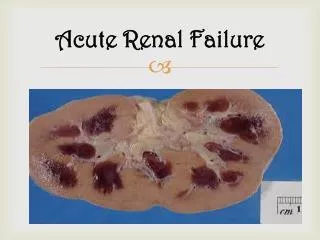
ARF- Renal • Useful to categorize according to Anatomical injury. • Primary sites, • Glomerulus- Acute Glomerulonephritis • Tubules- Acute Tubular Necrosis • Interstitium- Acute Interstial Nephritis • Vascular- Atheroembolism • ATN- most common • U/A-Protein, RBC,Casts,pigments
Acute Tubular Necrosis • Ischemic vs Nephrotoxic • Most frequently multi-factorial • Medical vs Surgical • Ischemic- Hypotension,shock • Nephrotoxic- Dye induced, Rhabdomyolysis
Acute Tubular Necrosis • Initiation, maintenance, recovery Phases • Mortality from very low to very high • Potentially Preventable • Long –term outcome in survivors very good
ATN- Specific Syndromes • Radiocontrast Nephropathy • Rhabdomyolysis • Aminoglycoside Related • Amphotericin B associated
Radiocontrast Nephropathy • 10% of Hospital acquired ATN • Mild and Transient in Majority • Risk factors, • Amount of Dye(> 100cc) • Volume Depletion • Renal Insufficiency • DM • Old Age • CHF • ACEI or NSAIDs
Radiocontrast Nephropathy • Risks higher with higher creatinine • Normal- negligible risks • Mild- Moderate RI(Creatinine< 2)– 5-10% risks • Mild- Moderate RI with DM- 10-40% risks • Advanced Renal Disease- >50%
Radiocontrast Nephropathy • Pathogenesis incompletely understood • Severe Renal vasoconstriction within seconds of contrast administration • Direct Renal Tubular injury • FENa < 1%
Radiocontrast Nephropathy • Independent risk factor of death • Prevention in high risk Patients • Consider Alternate imaging.g. MRI • Volume repletion with Saline • Minimize amount of Dye • Low Osmolality contrast media? • N-Acetylcysteine(Mucomyst)? • Fenoldopam-Selective Dopamine agonist? • Lasix, Mannitol, Dopamine –not helpful, may be risky • Prophylactic Hemodialysis- not helpful
Radiocontrast Nephropathy • N-Acetylcysteine – reducing agent, scavenge reactive oxygen species(ROS) • No good large randomized trial to prove its efficacy • Impact on morbidity and mortality unknown • Used commonly in practice b/o potential benefits and lack of Toxicity
Aminoglycoside Nephrotoxicity • Usually after 7-10 days • Depends on dose and frequency • Direct Proximal Tubular injury • Once a day dosing may be less Nephrotoxic • K. Ca. MG wasting • Risk factors- age, Renal insufficiency, Dose,Volume depletion
ARF from Rhabdomyolysis • Muscle injury leading to ARF • Most cases subclinical • Myoglobinuria cause, • Renal vasoconstriction • Proximal tubular damage • Intratubular cast (Obstruction) • Hypovolemia(Third Spacing) • Metabolic Acidosis, • Electrolyte Imbalance(K,Ca,P)
ARF from Rhabdomyolysis • Subclinical causes more common • Drugs • PVD • Seizure • FENa < 1% • U/A- Heme/+vie but no RBC • Aggressive Volume replacement • Urinary Alkalization?, Mannitol?
Amphotericin B Nephrotoxiciy • Very high incidence of ARF • Binds to sterol in cell membrane • Multiple sites in Nephrons • Distal Tubular Acidosis • Mg and K wasting • Dose dependent • Liposomal Amphotercin formulation less toxic • Saline loading helpful
Postoperative ARF • ARF after vascular,cardiac and major abdominal surgery. • Very high mortality • Multifactorial • 1-5% after CABG. • Risk factors, • Renal disease, cardiogenic shock,emergent surgery, Left main disease etc,
Acute Interstitial Nephritis • Classical triad(fever rash & eosinophilia) not usually seen • Mostly Drug related e.g. Cipro • Infection : Strept., Staph, CMV, EB virus, Hantaan virus etc • Systemic Diseases : SLE, Sarcoidosis. • Eosinophiluria may be absent • Dx by renal Biopsy. • Rx supportive, Hold Drug, Steroids ?
Atheroembolic ARF • Require high degree of suspicion • Cholesterol emboli • Renal failure – acute or subacute • Multisystem disorder • Lived reticularis • Digital Ischemia(Blue Toe Syndrome) • GI bleed, TIA, Rahbdomyolysis
Atheroembolic ARF • ARF after vascular procedure • ARF can be abrupt needing dialysis within few days. • Can be subacute occurring in staggered steps separated by stable renal function. • Patients on Anticoagulants are at high risk • Eosinphilia, eosinphiluria, low complement. • High mortality
Hepatorenal Syndrome • Profound renal vasoconstriction • Resemble Pre-renal Azotemia • Volume Expansion fail to improve renal function. • Pathogenesis incompletely understood • Oligiuric ARF, FENa low • Diagnosis of exclusion
Hepatorenal Syndrome • Two Types • Type 1 HRS: rapid ARF, hospitalized Pt.,>90% mortality • Type 11 HRS : insidious onset, slow progression of RI, refractory ascites, better prognosis. • ATN vs HRS • Low FENa I n ATN • casts in Bilirubinemia with HRS
Hepatorenal Syndrome • Rx difficult • Volume expansion with Albumin • Terlipressin(vasopressin analogue) • Midodrine (selective alpha 1 adrenergic agonist)+ octreotide(a somstoastatin analogue) • TIPS, Liver Transplantation • Dialysis in selective Patients
ARF in HIV/AIDS • Prerenal Azotemia • Renal salt wasting from Adrenal Insufficiency. • HIV Nephropathy • High risk for ATN • Drug side effects e.g. Pentamidine.Crystal nephropathy(indinavir) • TTP(prognosis worse ) • Rhabomyolysis
ARF from RPGN • Less common • Rapidly Progressive Glomerulonephritis include vasculitis, SLE, Wagner's • Active Urinary sediments(RBC cast diagnostic) • Higher degree of Proteinuria • Serology helpful(ANCA, ANA,IgMantibodyetc0 • Renal Biopsy usually required. • Early diagnosis essential to prevent ESRD • Rx with Steroids and Cytoxan
Rx of ARF • No proven Drugs • Many cause preventable • Volume expansion • Withdrawal of Drugs • Diuretics help in management but not curative • Dopamine potentially harmful
RRT in ARF • Renal Replacement Therapy usually the only option in severe ARF. • Indication of RRT • HYPERKALEMIA • METABOLIC Acidosis • Uremic Symptoms • Fluid Load • “Prophylactic” • RRT • Intermittent Hemodialysis • CVVHD • Extended Daily Dialysis(6-12h) • Peritoneal Dialysis- not favored
CVVHD vs Hemodialysis • HD – • more stable Pt, SBP >90, no heparin, allows larger amount of fluid removal in3-4 hours • CVVHD • Unstable Pt., low BP with high dose Pressers, allows gradual removal of fluids 24h • EDD • Allows no heparin dialysis, gradual removal of fluids, but expensive b/o Nursing Support
RRT- how to improve outcome? • Lot of Questions to answer • Frequency of Dialysis • Quantification of Dialysis • Type of Membrane of Dialysis • Synthetic vs. Cellulose • Does Erythropoietin improves outcome? • Faster fluid removal vs. slow fluid removal?