Download
1 / 33
1.4k likes | 7.84k Views
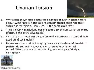
Ovarian Torsion. Stephanie Doniger, MD February, 2008. Ovarian Torsion. Twisting of the ovary on its pedicle 60% on right Compromising blood supply Ischemia: acute pain Surgical emergency! Restore perfusion, prevent necrosis. Ovarian Torsion. Incidence 2.7%

E N D
Ovarian Torsion • Stephanie Doniger, MD • February, 2008
Ovarian Torsion • Twisting of the ovary on its pedicle • 60% on right • Compromising blood supply • Ischemia: acute pain • Surgical emergency! • Restore perfusion, prevent necrosis
Ovarian Torsion • Incidence 2.7% • Rare in infants, children (17%) • Menarche • Cysts or masses (50-60%) • 4-6 cm • Pregnancy @ 4-16th weeks
Presentation • Mimics other causes of AP • Sudden onset severe pain • Focal, constant • Unilateral, lower abdominal/pelvic pain • Nausea, vomiting or urinary sx’s
Presentation • Intermittent exacerbations • Torse/detorse • PE • +/- painful mass @ abdomen or pelvis
Work-Up • Pregnancy test • Labs • Usu. not helpful • R/o other causes AP • Leukocytosis in children
Work-Up/Diagnosis • Color flow Doppler US • Visualize blood flow to ovary • CT: ovarian enlargement • Laparoscopic visualization
Prognosis • Salvage rates 10-27% • Up to 24-hrs after Sx onset • Complications • Infection, adhesions, • Chronic pain • Infertility
US: Questions • Abnormalities of the ovary • Ovarian cysts: ruptured, hemorrhagic • Ovarian torsion
US:Torsion • Ovarian enlargement • Mass, cyst • Unlikely w/normal ovarian size • Absent blood flow
US: Technique • Patient supine, thighs abducted • Endovaginal 5-7.5 MHz • Empty bladder • Transabdominal 3-5 MHz • Full bladder • Gray-scale • Doppler
Anatomy • Ovaries anterior & medial to iliac vessels • 2 x 2 x 3 cm • Pedicle • Broad ligament, fallopian tube • Uterine a & v, adnexal, uterine branches
Ovarian Pathology • Cysts • Hemorrhagic & simple • Masses • Follicular rupture • Complex fluid collections • Infection (TOA), ectopic
Sonographic Signs • Mass +/- pelvic fluid • Thickening of wall • Cystic hemorrhage • Twisted pedicle • Whirlpool, snail shell, target • Dilated veins
Sonographic Signs Whirlpool Sign Snailshell Vijayarghavan. J Ultrasound Med 23:1643-1649
Sonographic Signs Whirlpool Sign Vijayarghavan. J Ultrasound Med 23:1643-1649
Sonographic Signs Target Sign String of Beads Vijayarghavan. J Ultrasound Med 23:1643-1649
Doppler: Torsion • Absence of flow • Early or partial torsion • Arterial or venous flow can be maintained
Doppler: Torsion • False (-) • Early: arterial blood flow preserved • Impaired venous/lymphatic drainage • Period of de-torsing
Doppler: Torsion • False (+) • Salpingitis (tumor) • Endometriosis • Hemorrhagic cyst w/edema
Important Points • Risk factors for torsion • Important to scan in multiple planes • Normal Doppler imaging does NOT rule out torsion! • Clinical suspicion • Take the ovary seriously!
References • Baren JM, Rothrock SG, Brennan JA, Brown L. Pediatric Emergency Medicine. “Ovarian Torsion”. Elsevier 2008: 690-1. • Ma OJ, Mateer JR, Blaivas M. Emergency Ultrasound. “Gynecological Concepts: Ovarian Torsion.” McGraw-Hill 2008: 362. • Schraga ED, Kulkarni R, Blanda M. “Ovarian Torsion” eMedicine. Jan 29,2007. • Gaspari RJ, Fox JC, Sierzenski PR. Emergency Ultrasound: Principles and Practice “Ovarian scanning protocol”. Mosby 2006: 141-156 • Vijayaraghavan SB. Sonographic whirlpool sign in ovarian torsion. J Ultrasound Med. 2004. 23:1643-1649.