Download
1 / 33
330 likes | 484 Views
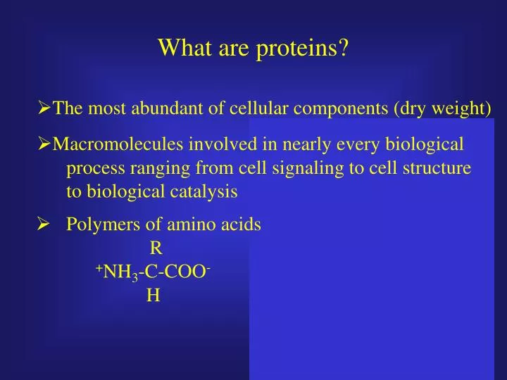
Macromolecular composition of E. coli. strain B/r grown. under a standard. culturecondition. (i.e., balanced growth,. Ø. Macromolecules involved in nearly every biological. glucose minimal medium, 37°C, mass doubling time of 40. min.):.

E N D
Macromolecular composition of E. coli strain B/r grown under a standard culturecondition (i.e., balanced growth, Ø Macromolecules involved in nearly every biological glucose minimal medium, 37°C, mass doubling time of 40 min.): process ranging from cell signaling to cell structure Macromolecules: to biological catalysis Protein 55% (of total dry weight) RNA 20.5% Ø Polymers of amino acids DNA 3.1% R Lipid 9.1% + NH - C - COO - Lipopolysaccharide 3.4% 3 Murein 2.5% H Glycogen 2.5% Soluble pool (amino acids, vitamins, etc.) 2.9% Inorganic ions 1.0% What are proteins? Ø The most abundant of cellular components (dry weight)
Basic formula for an amino acid NH2-C-COO- R H The properties of the amino acid depend on the side chain or R group
The net charge of an amino acid is influenced by pH NH2-C-COO R pKa = 9-10.0 pKa = 2.0 - +H H H pH = 12.0 pH = 7.0 pH = 1.0
The Henderson-Hasselbach equation relates pH to pKa Ka = [H+] [A-] [HA] [H+] = Ka[HA] [A-] HA H+ + A- -log[H+] = -log Ka + log [A-]/[HA] pH = pKa + log [unprotonated/protonated]
Review of the relationship between pH and amino acids
pH titration of amino acids determine their pKa values pI pKa = pH when [unprotonated/protonated] = 1
+NH3-CH-COO- +NH3-CH-COO- CH2 COO- CH2 CH2 COO- Acidic amino acids have a net negative charge at pH 7.0 Aspartic Acid (Asp; D) Glutamic Acid (Glu; E) pKa = 3.9 pKa = 4.3
Basic amino acids are positively charged at pH 7.0 Lysine (Lys; K) Arginine (Arg, R) +NH3-CH-COO- +NH3-CH-COO- (CH2)4 NH3+ (CH2)3 NH C NH2+ NH2 pKa = 10.8 pKa = 12.5
Other amino acids can be ionized under extreme pH conditions pH = 13 Serine (Ser; S) Tyrosine (Tyr; Y) +NH3-CH-COO- +NH3-CH-COO- CH2 OH CH2 OH - pKa = 13 Threonine (Thr; T) +NH3-CH-COO- CH CH3 OH - pKa = 10 - pKa = 13
Histidine is the only amino acid that is ionized within the physiological pH range Histidine (His; H) +NH3-CH-COO- • All other amino acids are • fully charged at pH 7.0 • while histidine can act • as a proton acceptor or • donor CH2 NH + N H pKa = 6.0 pH = 4.0 pH = 8.0
S S SH CH2 +NH3-CH-COO- Cysteine is a highly reactive amino acid Cysteine (Cys; C) • Sulfhydryl groups act as • cellular scavengers for • oxidants and xenobiotics • (ie. Glutathione) +NH3-CH-COO- CH2 SH • Formation of intra- and • intermolecular disulfide • bonds play an important • structural role in several • proteins pKa = 8.3
Asparagine and glutamine are uncharged polar amino acids Asparagine (Asn; N) Glutamine (Gln; Q) +NH3-CH-COO- +NH3-CH-COO- CH2 C O NH2 CH2 CH2 C O NH2 Hydrogen bond donor and acceptor
Hydrophobic residues are critical to protein structure Valine (Val; V) Phenylalanine (Phe; F) +NH3-CH-COO- +NH3-CH-COO- +NH3-CH-COO- CH CH3 CH3 CH2 Alanine (Ala; A) +NH3-CH-COO- CH3
Leucine (Leu; L) Isoleucine (Ile; I) +NH3-CH-COO- +NH3-CH-COO- CH2 CH CH3 CH3 CH CH3 CH2 CH3 Methionine (Met; M) +NH3-CH-COO- CH2 CH2 S CH3
Aromatic amino acids absorb near-UV light Tryptophan (Trp; W) +NH3-CH-COO- CH2 N H
The ring-like structure of proline restricts it’s flexibility in protein structures CH2 CH2 CH2 +NH2-CH-COO- Proline (Pro; P) Only amino acid that can form cis peptide bond
Glycine is the most “flexible” of the amino acids H +NH3-CH-COO- mass = 14 +24 + 32 + 5 = 75 amu or Da Molecular weight = 75 g/mole
- - Amino acids condense to form peptides Dipoles are crucial for protein structure and function
Amino acids other then glycine exist outside these parameters at an energetic cost
ab initio protein structure determination
Amino acids provide chemistry • Hydrophobicity (Non-polar residues) • van der Waal forces • Capacity for hydrogen bonds • Hydrophilic (Polar residues; electrostatic interactions) • pH sensitive ionization
Some amino acids are found more frequently in proteins Percent of total amino acids in entire ORF complement of genome
Chemistry important for protein structure • Hydrophobicity (Non-polar residues) Predominant force in protein folding • van der Waal forces Less than 1 kcal/mole • Capacity for hydrogen bonds bond length of 1.8 Angstroms, ~3-5 kcal/mole • Hydrophilic (Polar residues; electrostatic interactions) 4-7 kcal/mole • pH sensitive ionization (ie. disulfide bonds) • Although energy of covalent bonds is typically ~50-100 • kcal/mole, disulfide bonds contribute ~4 kcal/mole
There is great potential for hydrogen bonding in proteins
b-sheets form when two or more polypeptide chains are side by side antiparallel parallel Results from the trans orientation of backbone carbonyl and amine groups
“Regular” peptide structure results from amino acid interactions Helical secondary structure - • Classic alpha helix exhibits • H-bonds between backbone • carbonyl and amide groups • every fourth residue (i + 4) • p helix (i + 5) 310helix (i+3) – rare, at ends of alpha helices d i p o l e +
Domains are functional units of tertiary structure Elastase (1BMA)
Quaternary structures result from interactions between two or more proteins Maltoporin
Protein environment can have a dramatic effect on pKa’s of amino acid side chains The pKa of D26 of E. coli thioredoxin is ~7.5
Forces maintaining protein structure and things that disrupt them Ø Hydrophobicity (Non - polar residues) (disrupted by temperature, detergents, chaotropes, etc.) Ø van der Waal forces (easily disrupted) Ø Capacity for hydrogen bonds (also disrupted by pH and competitors such as urea) Ø Hydrophilic (Polar residues; electrostatic interactions) (can be shielded by salts and disrupted by pH) Ø pH sensitive ionization pH and oxidation-reduction reactions (DTT or BME)
To summarize: • Amino acids provide chemistry and • serve as the functional unit of higher order • peptides • Peptides use intramolecular interactions • to adopt secondary structures (a-helix, b-sheet) • Secondary structures fold into tertiary • “domain” structures • Some folded proteins interact to form • quaternary active structures