Download
1 / 32
320 likes | 382 Views
Understand the significance and causes of Microscopic Hematuria (MH) for proper evaluation and follow-up. Updated clinical guidelines and risk factors are highlighted. Guidelines provided for determination of MH, including the importance of urine specimen collection and associated risk factors for significant disease. Criteria for referral and potential underlying renal diseases are discussed.

E N D
Evaluation of Microscopic Hematuria Alon Z. Weizer, MD, MS Division of Urologic Oncology Department of Urology University of Michigan
Microscopic Hematuria (MH) • Objectives of Presentation • Appreciate the importance & possible etiologies of MH • Understand the proper determination of MH • Consider the steps in evaluation of MH • Articulate recommended follow-up in cases of MH with negative initial evaluation
Why Evaluate MH? • Gross hematuria is very often an indication of disease • Although less ominous, MH also can be an indication of disease • While many of these abnormalities are minor, some are significant and/or life-threatening
Possible Causes • Life-Threatening • Cancer • Renal parenchyma • Renal pelvic • Ureteral • Bladder • Prostatic • Urethral • Penile • AAA • Require Treatment • Calculi • Vesico-ureteralreflux • Infection • Ureteralobstruction • Symptomatic urethral obstruction • Renal parenchymaldisease • Symptomatic BPH • Renal artery stenosis • Renal vein thrombosis
Prevalence of MH • 0.2% to 16% in population studies • 2.5% to 21% in screening programs • 13% to 21% in men >60 years old • Likelihood of finding etiology of MH varies with risk • Low risk: Significant finding <5% • High risk: Significant finding >50%
Updated Clinical Guidelines • AUA Best Practices Panel: • Urology, Nephrology, Family Medicine, Radiology • Literature review & expert opinion • Critique of document by other physicians • Approval of AUA
Purpose of Recommendations • Resource for urologists & PCPs • Directed towards evaluation of asymptomatic MH in adults • Not meant to address: • Screening for hematuria • Gross hematuria • Symptomatic hematuria • Pediatric age group
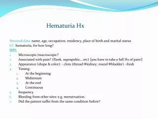
Determination of MH • Freshly voided, clean-catch, midstream specimen • Dipstick for Hb is screening test only • 95% sensitive, but only ~80% specific • In population with 10% hematuria, PPV of Dipstick is only 35% • Positive Dipstick must be confirmed by microscopy (>3 RBC/hpf)
Determination of MH • >3 RBC/hpf is standard criterion • 1 to 2 RBC/hpfdoesn’t require evaluation in most • However, it might if risk factors present • Just 1 specimen with hematuriashould prompt evaluation • Old rule: 2 of 3 properly collected specimens was standard criterion
Why the Change? • There is substantial evidence that MH caused by a serious underlying condition can be highly intermittent • Studies that evaluated patients after 1 positive sample, rates of malignancy in most were over 2% • Non-life-threatening diagnoses that would benefit from active management or follow-up are frequently found
Risk Factors for Significant Disease • Smoking history • Occupational exposure to chemicals or dyes • Benzenes or aromatic amines • History of analgesic abuse • Age >40 years • Significant urological history • Previous gross hematuria • Irritative voiding symptoms • Urinary tract infection • Prior pelvic radiation
Determination of MH • Hematuria cannot be determined in presence of squamous epithelial cells • These cells may indicate contamination from source outside urinary tract • Frequent in women • Difficultly obtaining perfect clean-catch specimen • Occasionally in men • Phimosis (cells from foreskin) • Squamous cells may be present in bladder urine
Determination of MH • If there appears to be hematuria, but squamous cells are present, then get catheterized specimen • Most common reason for “unnecessary” hematuria referral = contaminated specimen • Conversely, urine w/o RBCs is adequate for determination, even if there are squamous cells
Glomerular Hematuria • Origin of blood from renal parenchymal disease can be suggested by UA • Dysmorphic RBCs (variable size & shape w/irregular outline) → sensitive • Plain microscopy, or may need inverted phase-contrast microscopy
Asymptomatic MH History and Physical Examination Rule Out Benign Causes: Menstruation Vigorous Exercise Sexual Activity Viral Illness Trauma Infection Resolved after Tx or delay Persistent No Evaluation Evaluation Required
Evaluation: H&P • Gross or microscopic? • Gross more often assoc w/significant disease • Initial, terminal or total? • Initial = urethra, terminal = bladder neck/prostate • Pain, dysuria, bladder irritability • Suspect infection or obstruction • Anti-coagulated • Still require evaluation
Evaluation: H&P • Sickle cell, diabetes • Family or personal history of calculi, PCKD, other GU/Neph diseases • Trauma, physical or sexual activity • Tobacco use, occupational exposure • Association with menses • Fever, palpable mass, atrial fibrillation, visible blood at meatus, CVAT
Confirmed MH (obtain Serum Cr) Suggestion of Primary Renal Disease? Proteinuria* (> 500 to 1000 mg / day) Dysmorphic RBCs or RBC casts Elevated Serum Cr Yes No Urology Referral + Concurrent Nephrology Evaluation Urological Evaluation * Dipstick >1+ for protein prompts 24 hr urine
Purpose of Urological Eval • Upper tract imaging • Diseases of the kidney & ureter • Most commonly &/or significantly: • Calculi • Urothelial lesions • Obstruction • Renal masses • Cystoscopy • Diseases of the bladder & urethra • Most commonly &/or significantly: • Bladder cancer • BPH • Urethral strictures
Urological Evaluation for Asymptomatic MH Upper Tract Imaging Multi-phasic CT urography Alternative1 : MR urography Alternative 2: RPGs w/MRI Cystoscopy In patients ≥35 years & Patients w/RFs regardless of age (Discretionary: those <35 w/o RFs) Abnormal? Abnormal? Yes Yes Treat No No Consider Urine Cytology Follow-up Protocol
Imaging Modalities • Computed Tomography • Best modality for characterization of small renal masses • More widely available & less expensive than MRI • Best modality for calculi, renal and perirenal infection & associated complications • Sensitivity for urothelial lesions not defined with certainty, but thought to be good with “urography” technique
CT Urogram
Other Options • MR urography • Pregnancy, patients with iodinated contrast allergy • Risk of nephrogenic systemic fibrosis in patients with renal insufficiency • Retrograde pyelograms combined w/magnetic resonance imaging
Follow-Up Protocol • UA at 12 & 24 months • If negative for 2 consecutive years, then D/C • If persistent asympomatic MH, repeat urological evaluation w/in 3 – 5 years • If gross hematuria or new voiding sx, repeat urological evaluation
Before Urological Referral • Determine if this is real MH • >3 RBC/hpf • Catheterized urine if squamous cells • Exclude benign causes w/H&P • Likely glomerular → Refer to neph, too • Obtain serum Cr • Order imaging