Download

1 / 14
180 likes | 305 Views
Learn about osteomas, benign bone tumors primarily in the craniofacial skeleton, often asymptomatic in young adults. Different types like periosteal, endosteal, and cutis osteomas are discussed, with common locations and radiographic features detailed. Note the histopathologic features of compact and cancellous osteomas for accurate diagnosis and management.

E N D
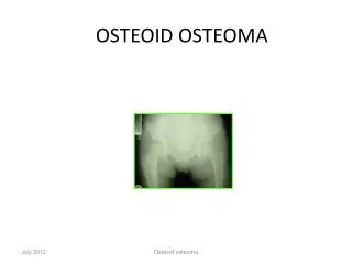
Osteoma benign neoplasms of bone forming tissue. Essentially restricted to the craniofacial skeleton Most in…… young adults generally asymptomatic
1. periosteal osteoma……a polypoid or sessile mass …… on the surface of the bone 2. endosteal osteoma……..in the medullary bone …… .. in the medullary bone 3.Osteoma cutis…….in the muscle or dermis The most common gnathic locations ………..bodyof the mandible Or the condyle. most osteomas occur posterior to the premolars on the lingual surface.
Condylar osteoma facial swelling pain limited mouth opening
Radiographically Circumscribed sclerotic masses Periosteal osteomas a uniform sclerotic pattern a sclerotic periphery with a central trabecular pattern or Smaller endosteal osteomas are difficult, if not impossible to differentiate from foci of sclerotic bone The true nature of these osteomas ................ continued growth.
Osteomas arising in the paranasal sinuses …….. sinusitis, headache, or ophthalmologic manifestations
Histopathologic Features 1.Compact osteomas normal-appearing dense bone showing minimal marrow tissue
Compact OsteomaCompact Lamellar bone Normal-appearing dense bone showing minimal marrow tissue
2.cancellous osteoma Trabeculae of bone and fibrifatty marrow
a much more cellular and active lesion with areas of loose vascular connective tissue containing irregular bone trabeculae. • Notice the considerable osteoblastic activity..
dense and lamellar cortical bone with a focal area of active bone modeling.