Download

1 / 41
410 likes | 448 Views
Diabetes foot workshop – Prevention and management of diabetic foot disease. Dr Samer Alsabbagh Dr Chantal Kong Dr Pawan Pusalkar Rebecca Gardner Caroline Leith Carolyn Wareham. Patient 1. 49 year old male Type 2 diabetes last 7 years

E N D
Diabetes foot workshop – Prevention and management of diabetic foot disease Dr SamerAlsabbagh Dr Chantal Kong Dr Pawan Pusalkar Rebecca Gardner Caroline Leith Carolyn Wareham
Patient 1 • 49 year old male • Type 2 diabetes last 7 years • Poor control and HbA1c 102 mmol/mol • Poor compliance to treatment, no diabetes follow-ups with GP • Painless sensory neuropathy
Presented to podiatry with 2 month hot, red, swollen left foot following a minor fall • Had 2 courses of antibiotics by GP • Good pedal pulses • Poor perception of sensations with 10g monofilament and tuning fork. • Left foot temp 4 degrees > right foot
Differential diagnosis • Further investigations & treatment
Foot X-ray – Features consistent with midfoot Charcots • Immobilisation in total contact cast offered but patient could not have it due to his personal circumstances • Aircast boot given • No change in clinical features after 2 weeks
Total contact cast applied • 3 weeks later developed a rub – planter aspect of 3/4th MTPJ 35 x 22mm. • Oral flucloxacillin for 2weeks • Cast removed and changed to aircast boot so that the ulcer could be monitored • Ulcer better in next 4-5 weeks only 5 x 5mm
Sudden worsening of the ulcer in the next appointment with swelling, redness and erythema of the whole foot • Unwell, fever, rigors last 24 hours • Had been walking a lot for last few days with the aircast and despite advice re walking to a minimum
Admitted as an emergency • MRI foot – Abscess at the planter aspect, oesteomyelitis of cuboid bone • Treated with IV antibiotics and discharged home on IV teicoplanin
8 months following diagnosis of Charcots – Rocker bottom foot deformity • Bony protrusion midfoot planter aspect • No planter ulcer • Wears removable bivalve plaster cast.
Patient 2 • 66 year old male with type 2 diabetes • Known Peripheral vascular disease and neuropathy • Admitted in an emergency under care of vascular surgeons in Dec 2015 with gangrene of left 2nd toe • Previous amputation of left 3rd, 4th and 5th metatarsals in 2007
Referred to foot clinic in April 2016 with an open necrotic ulcer on right 4th toe with exposed bone • Was already on antibiotics • Discussed in vascular MDT • Elective right anterior tibial angioplasty and amputation of right 4th toe performed
Learning Points High index of suspicion for Acute Charcots Refer early if in doubt Multidisciplinary team work improves outcomes
Epidemiology of Diabetic foot • Leading cause of all non traumatic lower limb amputation (40-60%) • Commonest cause of hospital bed occupancy (most common cause of hospital admission amongst diabetes patients, NaDIA) • 85% are preceded by foot ulceration • Lower limb amputations ↑x 15 in diabetes • > 50% require amputation of other limb within 3-5 years • 50% patients die within 5 years
1-4% of people with diabetes will develop an ulcer per year • Approximately 58% of DFU patients will become clinically infected. • The number of diabetes-related amputations in England has now reached an all-time high of 20 a day
Individuals with diabetic foot ulcers have a 50% chance of mortality in 5 years • Early diagnosis and early intervention by an MDT approach can achieve good outcomes
“Are diabetes-related wounds and amputations worse than cancer?”
Hospitalisation represents the greatest proportion of overall costs
Variation in amputation rates ‘shocking’ (BBC News 2010, 2012) • Department of Health data reveals the rate of major amputations in the South West, at 3 in 1000, almost twice the rate in the South East • Are we doing any better in Hertfordshire? • http://www.bbc.co.uk/news/health-17270379[17/08/2012 16:49:30] • http://www.bbc.co.uk/news/health-19050684[17/08/2012 16:48:14]7
Why the variation in foot outcomes? • What structures and systems are in place locally? (commissioning/organisation) • Are there clear guidelines/protocols for • -referral routes/care pathways? • -process of care? • Do patients & health care professionals know them, how and where to access them? • What are the current resources (podiatry, primary and secondary care,inpatients) • Are all patient being risk assessed appropriately? • Are all patients at risk being followed up as required? • Are the appropriate review/discharge care planning arrangements in place? (Continuity of care) • Optimal communication between HCPs involved (Use of different IT system)?
Quality Standards • Putting feet first 2003 • NICE NG19 August 2015, updated January 2016 Diabetes foot problems – Prevention and Management
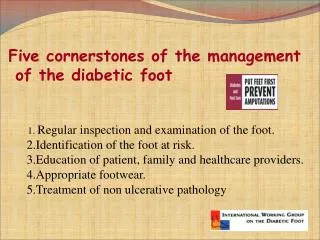
Reducing the risk of developing a diabetic foot problem • Education, Education, Education.…. (patients, carers, HCPs) • On-going care: annual review & recall • Detection of risk factors for ulceration • Classification of foot risk • Refer early to Community Podiatry & MDT foot clinics
How frequently should you assess your diabetic patients’ feet? • At time of diagnosis and at least annually thereafter • If any foot problems arise • On any admission to hospital, and if there is any change in their status while they are in hospital
Foot Risk stratification for the patient with diabetes (1) • Low risk (normal sensation, palpable pulses, +/-callus) • Moderate risk • High (increased) risk • Active & the Urgent/Emergency diabetic foot
Foot Risk stratification for the patient with diabetes (2) • Moderate risk • Deformity or • Neuropathy or • non-critical limb ischaemia • Review by Foot protection team (3-6 months) • High (Increased) risk • previous ulceration or • previous amputation or • on renal replacement therapy or • neuropathy and non-critical limb ischaemia together or • neuropathy in combination with callus and/or deformity or • non-critical limb ischaemia in combination with callus and/or deformity • Refer to Foot protection team (Community Podiatry). • Review 1-2 months, 1-2 weeks if any concern
Active diabetic foot problem • Ulceration or • spreading infection • Refer urgently to the MDT Foot clinic • If in doubt refer for admission • This is an emergency - Needs admission to hospital ●Critical limb ischaemia or • Gangrene or • Ulcer with fever or signs of sepsis ● Suspicion of an acute Charcot arthropathy, or an unexplained hot, red, swollen foot with or without pain
Management of active foot ulcers (1) • If clinical signs of active infection (redness, pain, swelling, discharge), give intensive systemic antibiotic therapy • -Flucloxacillin1g qdsPO for 7-14 days • -Doxycycline 100 mgs bdif penicillin allergic • Use wound dressings that best match clinical experience, patient preference, site of the wound (no strong evidence). Consider cost of dressing. • Regular monitoring & dressing change of wounds • Wound debridement (by specialist podiatrists or vascular surgeons) • Foot Xrayif wound persistent and deep with oedema, suspicious of osteomyelitis • Refer urgently to MDT Foot clinics and Community Podiatry • Admit if moderate to severe infection especially if patient septic or evidence of critical ischaemia
Management of diabetic foot ulcers (2) • For a diabetic foot ulcer to heal, the following conditions must be satisfied • Arterial inflow is adequate • Infection is treated appropriately • Pressure is removed from the wound and the immediate surrounding area • The most common cause of non-healing of neuropathic foot ulcers is the failure to remove pressure from the wound and immediate surrounding area
Diabetic Foot Osteomyelitis (DFO) • Diabetic foot Osteomyelitis is common problem, found in 20% overall to >60% (severe) diabetic foot infections –increase risk of LE amputation (up to 23) • Suspect if: • -long wound duration, recurrent infection • -wound deep >3 mm, large > 2 cm, bony prominence visible • -bone/joint -“sausage” toe • Probe to bone is useful if done and interpreted correctly • Blood tests: WBC, CRP, • -X-ray is the first test, limited sensitivity (early) & Specificity (late) -? -Repeat in 6-8 weeks if required • -if advanced imaging needed, MRI current best, marrow oedema
Management of Diabetic Foot Osteomyelitis (DFO) • Management is medical if detected and treated early • Infection typically contiguous spread from soft tissue • For wound culture, tissue specimens should be obtained by scraping the base of the ulcer with a scalpel or curette • Microbiology, pathophysiology: • S aureus predominant, Coagnegative Staph, • Beta-hemolytic streptococci (group A, B, and others) • Gram negative Rod (polymicrobial), • Anaerobic organisms • Rifampicin especially effective for bone infection; others Fluoroquinolones; Clindamycin; Daptomycin • RCT of DFO treated with antibiotics X 6 weeks Vs 12 weeks gave equivalent rates at 1 year
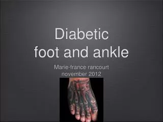
Acute Charcots foot • Condition affecting the bones, joints and soft tissue in the foot and ankle • Occurs in patients with diabetic neuropathy • Acute localised inflammation initially which leads to varying degrees and patterns of bone destruction, subluxation, dislocation and deformity • Despite neuropathy a lot of patients report of pain
Typical early clinical features are swollen, erythematous and hot foot • Early in the course often misdiagnosed as cellulitis, DVT or gout • Usually (but not always) peripheral circulation is preserved with patients having bounding pulses • Most common deformity is the rocker-bottom foot deformity
Investigations • Imaging – Foot X-ray should be the first imaging modality • Can show early changes but could be entirely normal despite clinical features of Charcots • Later on MRI foot or a nuclear medicine bone scan quite helpful to aid the diagnosis, MRI preferable in particular if there is an ulcer.
Treatment • Offloading in a total contact cast is the mainstay of treatment • Alternative is aircast boot • Offloading continued till temperature difference between feet is less than 2 degrees • Little evidence to support use of pharmacological therapies and we don’t use it • Surgical management – in a small subset of patients
An algorithm depicting the basic approach to the Charcot foot. *Osteomyelitis can be difficult to distinguish from the Charcot foot.
A Diabetic Foot Ulcer requires a team Diabetologist Paediatric Consultant The Patient Practice Nurse Diabetic Team Diabetes Specialist Nurse District Nurse G.P. Dieticians Tissue Viability Nurses Orthotist Podiatrists Pharmacist Orthopaedic Surgeon Radiologist Microbiologist Vascular Surgeon
How to refer to NHS Podiatry Tel 01582 711544 Fax 01582 765537 www.hct.nhs.uk/our-services/podiatry-service/ • Wound/Ulceration • Red, hot, swollen foot • Urgent fax to HCT Podiatry • HCT Podiatry does not provide home visits
When to refer to podiatry • Ongoing specialist foot car • Callus and corns in people at risk • Nail care for those at risk Hertfordshire Podiatry Service Harpenden Memorial Service Carlton Road Harpenden AL5 4TA Tel 01582 711544 www.hct.nhs.uk/our-services/podiatry-service/ Complete application form from HCT website and send to podiatry