Download
1 / 59
660 likes | 965 Views
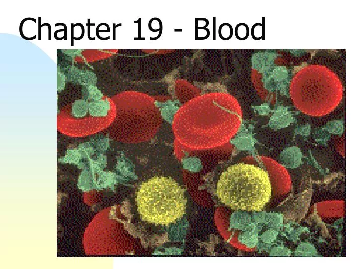
Chapter 19 - Blood. Functions of Blood. Transportation Delivery of nutrients, hormones Removal of wastes Regulation Body temperature pH Fluid volume Protection Prevent blood loss Prevent infection. Physical Characteristics of Blood. A liquid connective tissue A mixture

E N D
Functions of Blood • Transportation • Delivery of nutrients, hormones • Removal of wastes • Regulation • Body temperature • pH • Fluid volume • Protection • Prevent blood loss • Prevent infection
Physical Characteristics of Blood • A liquid connective tissue • A mixture • Formed elements - living blood cells • Plasma - fluid matrix • Heavier, thicker, more viscous than water • Due to ions, plasma proteins, blood cells • Composition and volume regulated by hormones • Temp - 38°C, higher than body temperature • pH - 7.4 (ranges from 7.35-7.45) • Volume differs between sexes, conditional • Female - average 4-5 L • Male - average 5-6 L
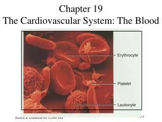
Components of Blood • Blood sample • Spin it • Separates into 2 parts • plasma • straw colored liquid on top • 55% of the volume • formed elements • solid portion on bottom • red blood cells • buffy coat - white blood cells and platelets • 45% of the volume • Hematocrit • percentage of formed element in a volume of blood • about 45% (higher in males than females)
Blood Plasma • Plasma • 92% water • 7% proteins • 1% other solutes
Blood Plasma • Proteins important (I ) for osmotic balance • Albumin • Fibrinogen (blood clotting) • Globulins • Regulatory proteins
Blood Plasma • Other solutes • Waste products • Nutrients • Regulatory substances • Gases • Electrolytes
Formed Elements • Formed elements • 99% red blood cells • 1% white blood cells and thrombocytes (platelets)
Formed Elements • Erythrocytes - Red Blood Cell's (RBC’s) IMPORTANT! Note the differences in relative size and appearance!
Formed Elements • Leukocytes - White Blood Cells • Granular leukocytes (granulocytes) • neutrophils • eosinophils • basophils • Agranular leukocytes (agranulocytes) • lymphocytes - T cells, B cells • Monocytes • Thrombocytes - platelets
Formation of Blood Cells • Hematopoiesis - blood cell formation • All blood cells come from pluripotent hematopoietic stem cells (hemocytoblasts) • Reside in red bone marrow • Give rise to precursor cells which develop into RBC’s, WBC’s and thrombocytes
Formation of Blood Cells • Erythropoiesis • RBC production, specifically • Hormonally controlled • Three phases • production of ribosomes • synthesis of hemoglobin • ejection of the nucleus and organelles • Leave bone marrow as reticulocyte • Mature in blood to a erythrocyte
Formation of Blood Cells • Regulation of RBC production • Regulated by negative feedback • O2 levels monitored in kidneys • hypoxia increases RBC production • Production stimulated by erythropoietin (EPO) from kidneys • Numbers • M - 5.4 million RBC’s/L (Testosterone stimulates EPO) • F - 4.8 million RBC's/L • 2 million cells released into blood/second • Anemia - Low O2 carrying capacity of blood • Insufficient # of RBC’s • hemorrhage - loss of RBC’s • hemolytic anemia - premature RBC rupture due to transfusion, disease, genetic problems • aplastic anemia • destruction or inhibition of hematopoietic components in bond marrow • toxins, drugs or irradiation • Decreased hemoglobin content • iron (heme) deficiency • insufficient iron due to diet or poor absorption
Formation of Blood Cells • Hematocrit • % of blood that is RBC’s • M 40-54% (47%), F 38-46% (42%), Why? • Indicates RBC production and hydration state • Abnormal Hct? • altitude – hypoxia, increased RBC’s • athletes - blood doping • anemia – decreased RBC’s • dehydration
Red Blood Cells (Erythrocytes) • RBC’s • 99% of formed elements • Function to carry O2 • Anatomy • Biconcave disks, 8µm in diameter • No nucleus or metabolic machinery • Filled with hemoglobin (Hgb) • O2 carrying protein • synthesized in cytosol before nucleus lost • 33% of cell weight
Red Blood Cells (Erythrocytes) • Normal Hgb in blood • Infants 14-20g Hgb/100ml • Adult • Male 14-15g Hgb /100ml • Female 12-15g Hgb /100 ml • 1.3 ml O2/g Hgb
RBC Physiology • RBC specialized for O2 carrying • 280 million Hgb molecules/cell • All internal space available for O2 carrying - no metabolism • Concave shape • Allows higher surface area/volume ratio increases gas diffusion • Allows passage through capillaries, very flexible
RBC Physiology • O2 combines with Hgb in lungs • O2 not very soluble in H2O • Need something to carry it • Hemoglobin • 4 globin (protein) chains - 2 chains, 2 chains • 4 non-protein heme pigments • Heme pigment has iron ion (Fe²+) that carries 1 O2 • Each RBC can carry about 1 billion O2 molecules • RBC's carry ¼ CO2 bound to hgb - forms carbaminohemoglobin
RBC Life Cycle • Life span • Only 100-120 days • Cells cannot repair damage due to loss of nucleus, ribosomes • Old RBC’s destroyed in liver, spleen • Macrophages eat old RBC's • Breakdown products recycled • Different pathways for each part of Hgb molecule • globin - AA's used for other protein synthesis • heme • iron portion - Fe2+ recycled • non-iron portion - bilirubin • released in bile, secreted into blood • bile enters intestine converted to urobilinogen by bacteria • gives urine/feces color
Granular Leukocytes Eosinophil Neutrophil Basophil
Neutrophil • 60-70% of all WBC’s • Anatomy • 10-12 µm diameter • 2-5 connected nuclear lobes • Fine, pale lilac granules • Physiology • Respond first to bacteria damage by chemotaxis • Phagocytosis • After engulfing pathogen release several chemicals • lysozymes • strong oxidants • defensins
Eosinophil • 2-4% of all WBC’s • Anatomy • 10-12 µm diameter • 2-3 connected nuclear lobes • red/orange large, uniform granules, do not block nucleus • Physiology • Exit capillaries, enter tissue fluid • Combat • parasites • histamine • Phagocytize antigen-antibody complexes
Basophil • 0.5-1% of all WBC’s • Anatomy • 8-10 µm diameter • Bilobed nucleus • Large granules round, blue-black, block nucleus • Physiology • Exit caps enter tissue fluids • Release heparin, histamine, serotonin – stimulate inflammation • Hypersensitivity (allergic) reactions
Agranular Leukocytes Lymphocyte Monocyte
Lymphocytes • 20-25% of all WBC’s • Anatomy • 7-15µm • Nucleus dark stained, round or indented • Cytoplasm sky blue rim around nucleus
Lymphocytes - Physiology • Immune response through lymphocytes responding to antigen • An antigen is: • Any chemical substance recognized as foreign when in body • Substance (mainly proteins) that stimulate immune response
Lymphocytes - Physiology • Two types of lymphocytes • B-cells • particularly active in killing bacteria • develop into plasma cells to form antibodies • antigen blockers that create antibody-antigen complex • complex prevents interactions w/ other body cells • T-cells • kill viruses, fungi, transplants, cancer, some bacteria • 3 types of cells • cytotoxic (killer) T cells - destroy foreign invaders • helper T cells - assist B cells and cytotoxic T cells • memory T cells - immune response memory
Monocytes • 3-8% of all WBC’s • Anatomy • 12-20 µm • Indented or kidney-shaped nucleus (not smooth) • cytoplasm foamy • Physiology • Slow to respond but arrive in larger numbers • Enlarge, differentiate into wandering macrophages • Clean up cellular debris, microbes following infection
Leukocyte Life Span and Number • Life span determined by foreign invaders! • Ingesting foreign bodies toxic, shortens cell life • Healthy WBC's - months/years, generally days • During infection may only live hours • fill with toxins • may die or burst
Leukocyte Life Span and Number • 5,000 - 10,000 WBC’s/mm3 blood • RBC/WBC ratio 700/1 • Differential WBC count (We have seen these before!) • Neutrophils 60-70% • Lymphocytes 20-25% • Monocytes 3-8% • Eosinophils 2-4% • Basophils 0.5-1%
Leukocyte Disorders • Leukopenia • Insufficient WBC production • Induced by drugs - glucocorticoids, anti-cancer drugs • Leukemia • Cancer (excess production) of the leukocytes • Bone marrow fills with cancerous (nonfunctional) leukocytes • Crowds out other cells types • anemia • bleeding
Leukocyte Disorders • Generally a descendent of a single cell • Different types of cells • myelocytic leukemia • lymphocytic leukemia • Under different cancerous conditions • acute - if derived from -blast type cells • chronic - if derived from later stages
QUIZ! Identify the marked cells! 1. 3. 2. Answers 1. Neutrophil 2. Basophil 3. Monocyte 4. Lymphocyte 5. Eosinophil 5. 4.
Platelets - Thrombocytes • Development • Megakaryoblasts shed small fragments • Each fragment has plasma membrane • Anatomy • 250,000-400,000/mm3 • No nucleus, disc shaped • 2-4 µm diameter w/ many granules
Platelets • Physiology • Short life span (5-9 days) • Help plug small holes in vessels • Granules several important functions • alpha granules • clotting factors • platelet derived growth factor (PDGF) • dense granules - Ca++, ADP, ATP, etc. • Also Thromboxane A2, vasoconstrictors, clot promoting enzymes
Hemostasis • 3 mechanisms in stopping bleeding • First - Vascular Spasm • Blood vessel constricts when damaged • wall smooth muscle contracts immediately • slows flow through vessel • reflexive?
Hemostasis • Second - Platelet Plug Formation • Platelet adhesion • platelets stick to exposed collagen • activates platelets • Platelet release reaction • platelets attach to other platelets • release granule contents (thromboxane A2) • promote vasoconstriction, platelet activation and aggregation • Platelet aggregation platelet plug • blocks blood loss in small vessels • not as good in larger vessels
Hemostasis • Third - Coagulation • Gel formation (clotting) in blood traps formed elements • Thrombosis - clotting in a normal vessel • Hemorrhage - slow clotting leading to a bleed
Hemostasis - Coagulation • A complicated process that works in a positive feedback cascade • 3 pathways - extrinsic, intrinsic and common • Pathways involve 12 factors in clot formation
Hemostasis - Coagulation • Overview
Hemostasis - Coagulation • Stage 1: Prothrombinase formation • Prothrombinase catalyzes Thrombin formation from Prothrombin • Stage 1 has 2 parts • Part 1: Extrinsic Pathway • Rapid (seconds) • Tissue factor (TF) enters blood from tissue • Ultimately activates prothrombinase Extrinsic Pathway Intrinsic Pathway Prothrombinase
Hemostasis - Coagulation • Stage 1: Prothrombinase formation (cont.) • Part 2: Intrinsic Pathway • Slower (minutes) • Activators in blood - damaged red blood or endothelial cells activate clotting • Extrinsic pathway also activates Intrinsic pathway • Ultimately activates prothrombinase • Ca2+ required for activation of both paths! Extrinsic Pathway Intrinsic Pathway Prothrombinase
Hemostasis - Coagulation • Stage 2 - Common Pathway • Thrombin Formation • requires enzyme Prothrombinase w/ Ca++ • catalyzes prothrombin conversion to thrombin • Thrombin accelerates formation of prothrombinase (positive feedback) • Thrombin accelerates platelet activation (positive feedback) + + Prothrombinase 2. Common Pathway
Hemostasis - Coagulation • Stage 3 - Common Pathway • Fibrin formation • activated enzyme thrombin w/ Ca++ • catalyzes fibrinogen fibrin • fibrinogen (soluble) • fibrin (insoluble) • Fibrin • Protein threads attach to vessel surface • Absorbs/inactivates 90% of thrombin, limits clot formation 3.
Hemostasis - Coagulation • Overview