Download
1 / 25
250 likes | 269 Views
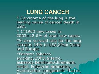
Lung cancer is a significant health concern with high mortality rates. Major risk factors include cigarette smoking and occupational exposure. The disease has various classifications based on tumor types and growth patterns. Clinical features vary depending on the disease stage, with late-stage symptoms including systemic and extrathoracic effects. Diagnosing lung cancer involves sputum cytology, biopsies, and imaging tests, while treatment options are limited due to operability rates and stagnant survival rates. Palliative care often becomes the primary approach. This overview aims to enhance understanding of lung cancer for better prevention and management.

E N D
Statistics Five leading causes of death in the early 2000s are: • Heart diseases; • Cancer; • Stroke; • Chronoc obstructive lung diseases; • Unintentional injuries.
Statistics Death rates, Adults, Ages 45-64 Whites Blacks Hispanics cancer cancer cancer heart dis. heart dis. heart dis. Injuries stroke cirrhosis COPD diabetes injuries Stroke injuries diaberes Diabetes HIV stroke
Statistics Lung cancer is the leading cause of cancer death in both sexes Male Female Lung - 31% Lung – 25% Prostate – 11% Breast – 11% ColonRectum–10% ColonRectum-11%
Statistics In 1998 in the US: 171,500 new cases of lung cancer 160,100 lung cancer deaths
Major Risk Factors • Cigarette smoking couses: a) loss of bronchial cilia b) basal epithelial hyperplasia c) nuclear hyperchromatism
Major Risk Factors • Occupational exposure • Air pollution a) sulfur dioxide b) carbonaceous particulate matter • Genetics a) p53 b) retinoblastoma gene
Lung Tumors Classification • 1. Benign neoplasms • 2. Bronchial carcinoids • 3. Bronchogenic carcinoma or lung cancer
Lung Benign Neoplasms • 1. Hamartomas • 2. Bronchial adenomas • 3. Leiomyomas • 4. Hemangiomas • 5. Lipomas • 6. Chondromas • 7. Teratomas • 8. Endometriosis
Lung (bronchial) carcinoids • The cells are related to the neuroendocrine argentaffin cell • The cells contain neurosecretory granules • The release of neuroendocrine substances leads to the carcinoid syndrome
Lung Cancer Classification Clinical and Anatomical Forms 1)Central cancer grows from a primary (main) , lobar, segmental bronchus; 2)Peripheral cancer grows from a subsegmental bronchus, bronchioles 3) Non-typical forms
Lung Cancer Classification Pathomorphological types of cancer: • Differentiated cancer a) adenocarcinoma b) squamous cell carcinoma c) bronchioalveolar carcinoma • Non-differentiated cancer a) small cells carcinoma b) large cell carcinoma
Lung Cancer Classification • adenocarcinoma (35%) Forms peripheral tumors from distal airways and alveoli Forms well-circumscribed gray-white masses that rarely cavitate Microscopically: a) well-differentiated b) poorly differentieted
Lung Cancer Classification • Squamous cell (25%) Arise from bronchial epitelium Starts as a small red granular plague Forms large intrabronchial mass Cavitation may occure Microscopically: intercellular bridges between neoplastic cells and keratin formation (“squamous pearls”)
Lung Cancer Classification • Bronchioalveolar carcinoma (5%) subset of adenocarcinoma that arise from terminal bronchioles or alveolar walls Forms peripheral nodules with mucinous gray translucence Microscopically: tall, columnar-cuboidal cells
Lung Cancer Classification • Small cell carcinoma (25%) Forms proximal large and soft gray-white massesthat can narrow bronchi circumferentially Microscopically:“oat-cell carcinoma” round or polygonal cells inconspicuous nucleoli modest amounts of cytoplasm
Lung Cancer Classification • Large cell carcinoma (15%) Forms peripheral lesions that become quite large and active Microscopically: large cells with citoplasm and distinct membranes, nucleoli and large nuclei No glandular differentiation Can form multinucleated giant cells
Lung Cancer Classification Depending on the grow character • Endobronchial cancer • Peribronchial cancer • Ramified (branching) cancer
Clinical Features: Two Modes Early stage: a) mild cough b) change of cough c) focal emphysema d) postobstructive atelectasis e) postobstructive pneumonia f) abscess formation
Clinical Features: Two Modes Late stage: • Nonspecific systemic symptoms • Intrathoracic symptoms • Extrathoracic extention symptoms • Paraneoplastic systemic syndromes
Clinical Features: Two Modes • Late stage: Paraneoplastic systemic syndromes 1)endocrine-methabolic syndromes 2) neuromuscular syndromes 3) hematologic/vascular syndromes 4) dermatoligic signs 5) skeletal and connective tissue syndromes
Clinical Features: Two Modes • Hormones secreted by tumor: 1) ACTH 2) MSH - skin pigmentation 3) PTH - hypercalcemia 4) ADH - hyponatremia 5) HCG - gynecomastia 6) Prolactin - lactation 7) Calcitonin - hypocalcemia 8) VIP – diarrhea, hypokalemia, achlorhydria
Diagnosis • 1. Sputum cytology • 2. Transbronchial biopsies • 3. Open biopsies • 4. Evaluation of liver • 5. Bone marrow biopsies
Treatment • Only 25 % of lung cancers are operable when discovered • 5-year survival rate has not improved over the last four decades • Palliation of symptoms becomes only the treatment for most patients