Download

1 / 56
580 likes | 761 Views
RECEPTORS. Receptors. Receptors are transducers which detect the Change in the environment (stimulus) & converts it into Propagated action Potential ( Impulse ). Classification of Receptors.

E N D
Receptors • Receptors are transducers which detect the Change in the environment (stimulus) & converts it into Propagated action Potential ( Impulse)
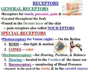
Classification of Receptors 1. On the basis of Modality of sensations Carried by Receptors ( Five Major Groups ) I. .Mechanoreceptors – ( Mechanical Change ) 1. skin tactile – Free Nerve Endings, Hair end organs etc 2.Deep tissues - ( Pacinian Corpuscles ) 3. Muscles / Tendons (Muscle spindles & Golgi tendons Organs 4. Hearing - ( Organ of Corti) 5. Equilibrium – ( Vestibular apparatus) 6. B.P. ( Baroreceptors ) II. Thermoreceptors - Cold & Warm Receptors III. Nociceptors (pain) - Free Nerve Endings
Classification of Receptors- contd • IV. Electromagnetic Receptors (Vision)- Rods & Cones. • V. CHEMORECPTORS; - Taste- Taste Buds - Smell – Olfactory Epithelium. - Arterial PO2- Aortic & Carotid Bodies - PCO2 - Central Chemo receptors - Osmolality - Neurons in Supraoptic Nucleus of Hypothalamus - Blood Glucose, A.A & F.A- Receptors in Hypothalamus
Classification of Receptors 2. On the Basis of Distribution in the Body i.e Somatic (Somesthetic) & Special Senses 3. On the basis of Origin of Stimuli: Interoceptors Proprioceptors Exteroceptors 4. Structural Classification Non Encapsulated Encapsulated 5. On the Basis of Adaptation: Phasic ( Rapidly Adapting)- Pacinian Corpuscles Tonic ( Slowly Adapting)- Pain (free Nerve endings)
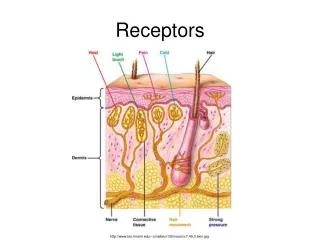
Mechanoreceptors Non-encapsulated receptors Skin tactile sensibilities (dermis & epidermis ) • Free Nerve Endings • Expanded Tip Endings Merkel’s Discs • Hair End Organs
1.Mechanoreceptors Un encapsulated Free NerveEndings Location: Widespread especially in Epithelia & Connective Tissue Modalities: touch, Pressure, Pain, Heat & Cold Nerve fiber: Type C
1.Mechanoreceptors Expanded Tip Endings-. Merkel’s Discs Location: Stratum Basale of Epidermis. Finger tips, Lips (Mostly Hairless Skin) Modality: Light Touch, Texture & shapes Informs for longer time Nerve type A-β
Hair End Organ Location:Entwined Around Hair Follicle Modality: Movement of Hair Movement of objects on the surface of the body Initial contact with the body Readily adapting. Nerve. A-β
Mechanoreceptors EncapsulatedEndings: . Meissner,s Corpuscles . Pacanian Corpuscles • Ruffini Corpuscles . Krause,s Corpuscles Meissner’s Corpuscl e Pacinian Corpuscle
Encapsulated Endings: Meissner,s Corpuscles.- Egg shaped Location: Dermal Papillae of finger tips, Palms, Lips, tongue, Eyelids Genitalia.(Hairless Skin only) Modality: Light Touch & Texture & low frequency vibration Adapts in fraction of seconds . Nerve A-β Particularly sensitive to movement of objects over the surface of the skin Meissner’s Corpuscl e
Encapsulated Endings Pacanian Corpuscles- Onion shape Location: Dermis, Joint Capsules & some Viscera Modality- Deep Pressure, Stretch & high frequency Vibration Adapts – Very rapidly Nerve A-β
Encapsulated Endings Ruffini Corpuscles. Multibranched encapsulated endings Location:Dermis, Subcutaneous Tissues & Joint Capsules Modality- Crude Touch & Pressure, Stretching of Skin, Joint movement Adapt slowly. Nerve. A-β Hence give information about continuous state of Deformation of the tissues.
EncapsulatedEndings Krause,s Corpuscles Minute cylindrical or oval bodies . Bulboid corpuscles Cutaneous thermoreceptors Conjunctiva of eye Mucous membrane of lips and tongue Penis and clitoris
Mechanoreceptors • Deep TissuesSensibilities: Free Nerve Endings Expanded Tip Endings- Merkle’s Discs Spray endings- Ruffini’s Endings • Muscles- Muscles Spindles, &Golgi Tendon organs. • Hearing - Organ Of Corti • Equilibrium- Vestibular Apparatus • B. Pressure - Baroreceptors
Transmission in Peripheral Nerve fibers Type Aβ nerve fibers (30-70m/sec) Meissners corpuscles Iggo dome receptors Hair receptors Pacinian corpuscle Ruffinis endings.
Transmission in Peripheral Nerve fibers Free nerve endings : Pain Type Aδ myelinated (5-30m/sec) Type C un myelinated fibers ( 0.5-2m/sec) All tactile receptors detect vibration although different receptors detect different frequencies.
Proprioception(Sense of Position) • Static Proprioception. Conscious perception of the orientation of the different parts of the body with respect to each other • Dynamic-Conscious perception of rate of movement ( Kinesthesia)
Multiple Receptors are involved in Proprioception. Muscle Spindles (important) Pacinian Corpuscles other tactile receptors also participate
Tactile Receptors in the Skin Figure 15.3a-f
Doctrine of Specific Nerve Energies The specificity of a sensory receptor for a particular type of stimulus is called the law of specific nerve energies.
Law of Projection: Conscious perception of a sensation produced is always referred to the location of the receptor, no matter where a sensory pathway is stimulated along its course to the cortex. Phantom limb
Phantom limb • Neuromas. The ends of the nerves cut at the time of amputation often form nerve tangles called neuromas.
Phantom limb Cortical Plasticity. • Brains remarkable ability to reorganize itself by forming new neuronal connections based on individual experiences, life style and environment. • In brain neuronal connections and cortical maps are continuously remodeled through out life.
Topographical MAP Broadman’s ares
Sensory Transduction • Q:How is a stimulus converted into a neural signal? • Ans: • The stimulus opens ion channels in the receptor membrane, either directly or indirectly (through a second messenger). In most cases, channel opening results in depolarization of the membrane .(Excitation) • In a few cases, the response to the stimulus is Hyperpolarization when K+ leaves the cell or Cl- enter the cell. ( Inhibition)
Sensory Transduction • Sensory transduction converts stimuli into graded potentials or Receptor potential • MECHANISM: 1. By Mechanical Deformation 2. By application of Chemical (Neurotransmitter) 3. By change of Temperature 4. By electromagnetic radiation e.g light
Sensory Transduction. Contd. Receptor or Generator Potential; • When Receptor Potential rises above the threshold level ,action potential is elicited in the nerve attached to the Receptor. • Frequency of A.P > more the Receptor Potential above the threshold level
Sensory Representations • To create an accurate neural representation of sensory stimuli, the brain must distinguish FOUR properties of stimulus : • 1)stimulus modality • 2) stimuluslocation • 3)stimulus intensity • 4) stimulus duration
Stimulus Modality • Each receptor type is most sensitive to a particular type of stimulus (Modality). The brain thus associates a signal coming from a specific group of receptors via specific nerve fibers with a specific modality. • This specificity of nerve fiber for transmitting only one modality of sensation is called labeled line Principal.
Stimulus Location • The area of the body that when stimulated leads to activity in a particular afferent neuron is called the receptive field for that neuron • When action potentials are elicited from a sensory neuron, the neuron’s receptive field codes the stimulus location.
Stimulus Location • Sensory receptive fields vary in size and frequently overlap.
Stimulus Location • Lateral inhibitionenhances the contrast between the stimulus and its surrounding, facilitating its perception and localization.
Stimulus Location • Sensory neuronal receptive fields are orderly organized in cortical sensory areas to form topographical maps. • The location of a stimulus is coded according to which group of neurons are activated
Topographical MAP Broadman’s ares
Stimulus Location (Exception) • Auditory information is the exception to the topographical localization rule. • For this sensory modality the brain uses the timing differencein receptor activation to compute the source location of sounds
Locating sensations from internal organs is less precise than from the skin because there are few afferent neurons in the internal organs and each has a larger receptive field.
Vision hearing and smell stimulus location is interpreted as arising from the site from which the stimulus originated than the place on our body where the stimulus is actually applied.
Stimulus Intensity • Stimulus intensity is transmitted to the brain by two mechanisms: • 1) the number of receptors activated (population coding), from low-threshold receptors to high-threshold ones. • 2) the frequency of action potentials (frequency coding), following not a linear but a power relationship. (Weber-Fechner Law)
Weber-Fechner- Law • During the stimulation of a receptor, if the response given by the receptor is to be doubled the strength of stimulus must be increased 100 times.