Download
1 / 105
1.06k likes | 1.14k Views
Comprehensive module covering pathophysiology of angina, acute myocardial infarction, and stroke, and cardiovascular anatomy. Learn field assessment, EKG rhythms, and medication dosing, with emphasis on ACS development and risk factors.

E N D
Angina, Acute MI, & Acute Stroke August 2014 CE Condell Medical Center EMS System Prepared by: Sharon Hopkins, RN, EMT-P, BSN Rev 8.18.14
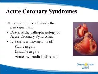
Objectives • Upon successful completion of this module, the EMS provider will be able to: • 1.Describe the pathophysiology of angina. • 2. Describe the pathophysiology of the acute myocardial infarction process. • 3. Describe the atypical presentations of women, elderly, and those with long standing diabetes.
Objectives cont’d • 4. Describe the pathophysiology of ischemic and hemorrhagic strokes. • 5.Describe field assessment of the patient with a possible stroke including documentation of time of onset, blood sugar level, and Cincinnati Stroke Scale. • 6. Actively participate in review of selected Region X SOP’s.
Objectives cont’d • 7. Actively participate in review of a variety of EKG rhythms and 12 lead EKG’s. • 8. Actively participate in case scenario discussion. • 9. Actively participate in calculating and preparing medication doses for the pediatric patient. • 10. Review responsibilities of the preceptor role. • 11. Successfully complete the post quiz with a score of 80% or better
Cardiovascular Anatomy • Cardiovascular system has 2 major components • Heart • 4 chambered pump • 2/3 of mass is to left of sternum • Apex just above diaphragm • Base, top of heart, lies at level of 2nd rib • Peripheral blood vessels • Transport system to deliver blood to the body and to transport waste for removal
Layers of the Heart • Pericardium – protective sac around heart • Epicardium - Outer most layer • Coronary vessels lie on this epicardial layer • Myocardium • Thick middle layer of cells with electrical properties • Endocardium - Inner most layer • Lines heart chambers; in contact with blood flow within the chambers
Cardiac Damage Post MI • Each individual is unique • Damage may be only on the surface or penetrate all layers of the heart • The greater the level/degree of damage to the heart the more negatively affected the heart is to work as a pump
What is Acute Coronary Syndrome (ACS)? • A list of diagnoses that affect the cardiac system • Indicates an interruption of blood flow to the heart • Unstable angina • Non-ST elevation MI • ST elevation MI
What Causes ACS To Develop? • An imbalance between supply and demand of O2 • Atherosclerotic plaque rupture in coronary artery • Thrombosis (clot) formation in artery • Coronary spasm • Dissection of blood vessel • Increased demand of O2 in face of fixed obstruction
Main Contributing Risk Factors to Cardiac Problems • Hypertension • Hyperlipidemia • Smoking • Diabetes • Notice: all of the above are considered modifiable risk factors – you can do something to control them!
Pathophysiology of angina • Chest pain/discomfort related to a decrease in oxygen-rich blood flow • Usually due to coronary artery disease (CAD) • Atherosclerosis • Build up of plaque over time that narrows the internal diameter of the vessel • Arteriosclerosis • Stiffening of vessels over a period of time which makes them less pliable
Pathophysiology of Angina • Plaque formation • Angina serves as a warning that something is going on
Stable Angina • Most common form • Pain occurs when oxygen demand is greater than the supply during periods of increased workload of the heart • Can usually predict activities that will trigger an event • Usually treated with rest and medication (i.e.: nitroglycerin) • This is a warning that the patient may have an acute MI in the future
Unstable Angina • Pain that is unpredictable and can occur at rest • May not stop with rest and/or medication • Event to be taken seriously • May be predicting an imminent acute MI in the near future
Variant Angina • Occurs when vessel is in spasm • Very painful • Often occurs at night • Controlled with medication
Treating Angina • Treated as if the patient were experiencing an acute MI • Patient should stop activity and rest • Question if the patient has taken their own nitroglycerin or other medication • Often, if the pain is relieved with rest and nitroglycerin, it is usually angina, not acute MI
Pathophysiology of Acute MI Process • Significant blockage of one or more coronary arteries • Atherosclerosis is gradual process, typically over decades, of plaque build-up on arterial walls • Size and area affected impacts outcome • Location, location, location!
Ischemic Cascade • Heart cells around blocked coronary artery die • These do not regenerate • Collagen scar forms • Scarring increases risks for potentially life-threatening dysrhythmias • Weakened muscle walls vulnerable to formation of ventricular aneurysm which could rupture at any time • Injured heart tissue conducts electrical impulses more slowly • Allows for possible development of re-entry or feedback loop that can generate dysrhythmia
Pathological Types of Acute MI • Transmural – atherosclerosis affected a major coronary artery • MI extends through whole thickness of heart muscle • Anterior, posterior, inferior, lateral, septal walls • ST elevation noted on EKG and Q waves develop • Subendocardial • Small area below surface of wall involved in left ventricle, ventricular septum, or papillary muscle • ST depression noted on EKG
Coronary Circulation • Coronary arteries (CA) • Vessels that originate in aorta • Supply the heart with it’s blood flow • Main CA lies on surface of heart – epicardial coronary arteries • Small penetrating arteries supply myocardial muscle
Collateral Circulation • Protective mechanism to provide alternative path for blood flow in case of system blockage • Takes time to develop
Coronary Arteries • Left coronary artery • Left ventricle • Interventricular septum • Part of right ventricle • Heart’s conduction system • 2 branches • Anterior descending artery (LAD) • Circumflex artery
Coronary Arteries cont’d • Right coronary artery • Portion right atrium, right ventricle, and part of conduction system • 2 branches • posterior descending artery • Marginal artery
STEMI Waveform Pattern • Evaluation of ST segment elevation • 0.04 seconds after J point • >2mm for males in V2 and V3 • >1.5mm females in V2 and V3 • >1mm in other EKG leads • Early STEMI may just display peaked T wave • ST elevation develops later
Myocardium and Vessel Structure • Notice that coronary arteries lie on epicardial surface
Patient Complaints and Evaluation • Symptoms that persist beyond 15 minutes • Remember that symptoms may be vague • Always consider worse case scenario and hope for the best • When in doubt, obtain a 12 lead EKG • Remember: a negative 12 lead EKG does not mean the patient is NOT having an MI • We are just positive when ST elevation is present
Mimics of Chest Pain • Occurrences of chest pain must be fully evaluated • Assume the worst; hope for the best • Need to consider other differential diagnosis putting the one that is most life threatening at the top of the list • Remember to not be fooled by atypical presentation of AMI
Differential Diagnosis…What Could This Be??? • Stable angina • Acute pericarditis • Aortic dissection • Pulmonary embolism • Pneumonia • Pneumothorax • Esophageal spasm • Reflux • Gastritis • Cholecystitis • Pancreatitis • Musculoskeletal pain
Non-atherosclerotic Causes AMI in Younger Patients • Emboli from infected cardiac valves • Coronary occlusion from vasculitis • Coronary artery spasms • Cocaine use • Congenital coronary anomalies • Coronary trauma • Increased O2 requirement • Decreased O2 delivery (i.e.: anemia)
Typical Patterns of Chest PainMay be from surge of catecholamines in response to pain and hemodynamic abnormalities from cardiac dysfunction
Atypical Presentations • Women, elderly, long standing diabetics • Back pain • Jaw pain • Right arm pain • Indigestion • Nausea • Fatigue/weakness • Dyspnea/ shortness of breath • No pain
Atypical Presentation in Women • Most frequent complaints in women are shortness of breath, weakness, feeling of indigestion, and fatigue
Women and Acute MI • What causes atypical presentations? • Anatomical factors • Physiological factors • Pathological factors • The above list may demonstrate the causes of the differences between males and females in typical versus atypical presentation of acute MI
Atypical Presentations of Elderly • Most likely complaints • Dyspnea • Diaphoresis • Nausea • Syncope • May have atypical presentation most likely due to altered pain perceptions
Atypical Presentation of Long Standing Diabetes • Silent ischemia considered due to autonomic denervation of heart • Nerve damage that disrupts signals between the brain and other body parts • Diabetics recognized at risk for atherosclerotic plaque formation and thrombosis contributing to acute MI
Diagnosing Cardiac Problems • Patient history of events • 12 lead EKG analysis – changes over hours & days • Large peaked T waves, then ST elevation, then negative T waves, then pathological Q wave development • Cardiac markers – blood work • Timing of the rise and fall of enzymes can help predict when the acute process occurred • Troponin I levels • Specific to myocardial damage
Have you ever wondered… • What will I see on the EKG if the patient is having angina and not an MI? • ST elevation can occur (rarely) if perfusion is poor or some spasm is present; it could take some time for the ischemia to normalize • When ST elevation on EKG is from a paced rhythm or LBBB, how do you decide when to take the patient to the cath lab? • Most often history and how the patient looks are deciding factors. Cardiologists would rather find normal arteries than miss an MI. Comparison with old EKG’s could help if they are available
Questions cont’d…. • If the patient is having atypical angina attacks, what is the likelihood that they will have an acute MI? • This is very predictive with a rate of at least 30% within 30 days • Do all persons with an MI develop a diagnostic Q wave that shows on the EKG forever? • Diagnostic Q waves do not develop immediately and may not if response and repair is rapid. They depend on the thickness of the infarct and length of time before treatment
Delay in Seeking Care • Atypical presentations (absence of chest pain) increase the delay before seeking care • TIME IS MUSCLE!!! • Delay in appropriate assessment and intervention increases the mortality rate
What Therapies Are Available for Diagnosing and Treating AMI? • Angiogram / Cardiac catherization • Insertion of long flexible tube into artery or vein • Die injected to evaluate blood flow through coronary arteries • Fibrinolytic therapy • Medication used to dissolve the clot
Therapies cont’d • Angioplasty / PTCA • Percutaneous transluminal coronary angioplasty • Balloon tipped catheter introduced into femoral artery • Advanced to coronary artery • Balloon expanded to press clot into wall of artery • Stent may be left to keep lumen open • “Door-to-balloon” phrase often heard
Therapies cont’d • CABG / “open heart” • Coronary artery by-pass graft surgery • Small vein removed from the patient (i.e.: leg) and attached to the aorta and then a point beyond and by-passing the blockage
LBBB Pattern & Paced Rhythms in Acute MI • Diagnosis difficult based on the EKG • The baseline ST segments and T waves shift masking or mimicking acute MI • Assume acute MI and treat as such • Hospital assessment includes lab analysis and MD assessment • Patients may be taken to the cath lab on presumptions • MD would rather err on side of being aggressive than to be delayed in treatment of acute process