Download
1 / 20
200 likes | 224 Views
Dive into the intricacies of muscle contractility, from the structure of skeletal muscle to the molecular basis of contraction and energetics of filament sliding. Explore key components like actin, myosins, and sarcomeres.

E N D
9.6 Muscle Contractility (1) • A skeletal muscle fiber is a multinucleate cell as a result of fusion of myoblasts in the embryo. • A single muscle fiber is large and highly organized. • Each muscle fiber contains hundreds of cylindrical strands called myofibrils.
Muscle Contractility (2) • Each myofibril consists of a repeating array of sarcomeres. • Each sarcomere has a banding pattern that gives muscle fiber a striated appearance. • Banding pattern: • Thin filaments (I and A bands) • Thick filaments (H and A bands)
Muscle Contractility (3) • The sliding Filament Model of Muscle Contraction • Skeletal muscle works by shortening fibers. • A bands remain constant in length. • H and I bands decrease in width. • Z lines on both ends of sarcomere move inward.
Muscle Contractility (4) • The Composition and Organization of Thick and Thin Filaments • Thin filaments contain actin as well as tropomyosin and troponin. • Tropomyosin occupies the gap between two actin molecules. • Troponin molecules are in contact with both actin and tropomyosin.
Muscle Contractility (5) • The third most abundant protein of skeletal muscles is titin. • Titin filaments are elastic and prevent the sarcomere from being pulled apart during muscle stretching.
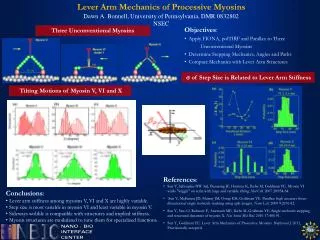
Muscle Contractility (6) • The Molecular Basis of Contraction • During contraction, myosin heads bend thus sliding the thin filaments over the thick filament. • Energy released from ATP hydrolysis induces a conformational change within the head. • Elongated myosin neck acts as a “lever arm”. • Attached actin filament slides a much greater distance than would be possible.
Muscle Contractility (7) • The Energetics of Filament Sliding • Energy is provided by ATPase activity in the myosin head. • Activated myosin attaches to actin initiating the power stroke. • Release of bound ADP is followed by binding of another ATP. • Absence of ATP prevents dissociation of cross-bridges causing rigor mortis.
Muscle Contractility (8) • Excitation-Contraction Coupling • Contact between nerve and muscle is called the neuromuscular junction. • The linking of the nerve impulse to the shortening of the sarcomere is referred to as excitation-contraction coupling. • Action potential in muscles is propagated into the cell interior by transverse (T) tubules.
Muscle Contractility (9) • Excitation-contraction coupling (continued) • T tubules terminate near the sarcoplasmic reticulum (SR), which stores Ca2+. • In a relaxed sarcomere, Ca2+ levels are low. • An action potential opens calcium channels in the SR, releasing Ca2+. • Binding of Ca2+ to troponin causes a conformational changes, shifting tropomyosin and exposing the myosin binding site.