Download
1 / 63
640 likes | 674 Views
Discover the synthesis and structure of major glycan classes including N-linked, O-linked, and glycolipid oligosaccharides. Explore processing in the ER and Golgi, and evolutionary variations in glycan pathways. Learn about N-glycan and O-glycan structures in a cellular context.

E N D
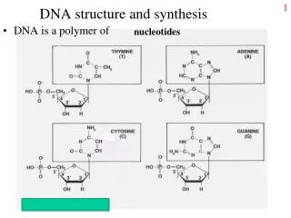
Large O-linked Glycosaminoglycans and poly-lactosamine structures • Glycoprotein N-linked and O-linked oligosaccharides • Glycolipid oligosaccharides
Symbolic Representation Simplified Traditional The building blocks After Varki, A
Glycan synthesis in a cellular context
Overview From ER through Trans-Golgi and points inbetween
“Hybrid” Major Classes of N-Glycans “Complex” “High-Mannose” (oligo-mannose) After Varki, A
GlcNAc Man Glc Gal Sia Fuc Dolichol Biosynthesis of N-Glycans:Production of GlcNAc-P-P-Dolichol Tunicamycin Blocks - not very specific! Adapted from Marquardt T, Denecke J. Eur J Pediatr. 2003 Jun;162(6):359-79
GlcNAc Man Glc Gal Sia Fuc Biosynthesis of the N-GlycanPrecursor on the Cytosolic Leaflet of the Endoplasmic Reticulum (ER) CDG = Congenital Disorder of Glycosylation in Humans Adapted from Marquardt T, Denecke J. Eur J Pediatr. 2003 Jun;162(6):359-79
GlcNAc Man Glc Gal Sia Fuc Biosynthesis of the N-GlycanPrecursor on Lumenal Leaflet of ER Adapted from Marquardt T, Denecke J. Eur J Pediatr. 2003 Jun;162(6):359-79
GlcNAc Man Glc Gal Sia Fuc Completion of Biosynthesis of N-GlycanPrecursor on Lumenal Leaflet of ER- and Transfer to Protein Adapted from Marquardt T, Denecke J. Eur J Pediatr. 2003 Jun;162(6):359-79
Target “sequon” for N-glycosylation • Necessary but not sufficient • X = any amino acid except proline • Rarely can be Asn-X-Cys • Transfer co-translational/immediate • post-translational before folding • ~2/3 of proteins have sequons • ~ 2/3 sequons actually occupied • (some variably) Yeast OST complex contains nine membrane-bound subunits Oligosaccharyltransferase complex (OST) in the ER membrane transfers the dolichol N-glycan precursor to asparagine residues on nascently translated proteins After Varki, A
GlcNAc Man Glc Gal Sia Fuc Initial Processing of N-Glycans in the ER and Golgi ER Golgi Adapted from Marquardt T, Denecke J. Eur J Pediatr. 2003 Jun;162(6):359-79
Improperly folded proteins are re-glucosylated by glucosyltransferase which acts as “sensor” for improper folding 3 Glucose Residues Calnexin (and Calcireticulin) function during glycoprotein folding in the endoplasmic reticulum After Varki, A
Basic Glycosylphosphatidylinositol (GPI) Anchor Phospholipid After Hart, G
Cell surface hydrolases Protozoal antigens alkaline phosphatase trypanosome VSG acetylcholinesterase leishmanial protease 5’ nucleotidase plasmodium antigens Adhesion molecules Mammalian antigens neural cell adhesion molecule carcinoembryonic antigen heparan sulfate proteoglycan Thy-1 Others scrapie prion protein folate receptor decay accelerating factor Examples of GPI-Anchored Proteins After Hart, G
Structure of the Basic GPI Anchor After Hart, G
Structural Analysis of the GPI AnchorEnzymatic and chemical cleavage sites are useful in identifying GPI anchored membrane proteins After Hart, G
Examples of C-Terminal Sequences Signaling the Addition of GPI-Anchors Bold AA is site of GPI attachment Sequence to right is cleaved by the transpeptidase upon Anchor addition After Hart, G
GlcNAc Man Glc Gal Sia Fuc Completion of Processing of N-Glycans in ER and Golgi Final products often show “microheterogeneity” at each N-Glycosylation site Adapted from Marquardt T, Denecke J. Eur J Pediatr. 2003 Jun;162(6):359-79
GlcNAc-Transferases Determine Number of“Antennae” of N-glycans After Varki, A
Some representative examples of mammalian complex-type N-glycans After Varki, A
Slime Yeast Plants Mold Insects 3 a a3 6a a3 6a a 4 N N Asn Asn a 4 b 2 a 4 a3 a6 N N Vertebrates Asn Asn Evolutionary Variations of the N-glycan Processing Pathway “Pauci- mannose” After Varki, A
O-Glycosidic Linkage O-glycosidic linkage is sensitive to alkali (regardless of stereochemistry) b-elimination GalNAc a Ser After Esko, J
Man Ser/Thr Ser/Thr Ser/Thr Ser/Thr Examples of O-Glycans Yeast mannoproteins a-dystroglycan Nuclear Proteins Cytoplasmic Proteins GalNAc Fuc GlcNAc Mucins Notch Coagulation Factors Fibrinolytic Factors After Esko, J
a3 b4 a3 b3 b4 b3 b4 a3 b3 b6 Ser/Thr Mucin-Type O-GalNAc Glycans • Major vertebrate O-glycan • Begins in cis-Golgi by attachment of GalNAc in a-linkage to specific Ser/Thr residues • Assembly is simpler than N-linked chains - no lipid intermediate is used • Always involves nucleotide sugars • Always occurs by addition to non-reducing terminus or by branching a After Esko, J
Polypeptide GalNAc Transferases • 12 members of mammalian ppGalNAcT family • Estimated size of family = 24 • Share structural features in active site • Some have lectin (ricin) domain Regions in white, pink, red, and black represent, respectively, 0–29%, 30–69%, 70–99%, and 100% sequence identity (Hagen et al. (2003) Glycobiology 13:1R-16R). After Esko, J
b3 b6 b3 Ser/Thr Ser/Thr Ser/Thr Core 1 and Core 2 Synthesis Core 1 GalT Core 2 GlcNAcT After Esko, J
Ser/Thr Core 3 and Core 4 Synthesis Core 3 GlcNAcT Core 4 GlcNAcT b3 b3 b6 Ser/Thr Ser/Thr After Esko, J
Core 1 Core 2 Core 3 Core 4 b3 b6 Ser/Thr Core 6? Core 7 Core 8 Core 5 b6 a6 a3 b3 a3 b3 b3 b6 Ser/Thr Ser/Thr Ser/Thr Ser/Thr Ser/Thr Ser/Thr Ser/Thr Unusual Core O-Glycan Structures After Esko, J
Mucins are Heavily O-glycosylated • Apomucin contain tandem repeats (8-169 amino acids) rich in proline, threonine, and serine (PTS domains) • Glycosylation constitutes as much as 80% of mass and tend clustered - bottle brush • Expressed by epithelial cells that line the gastrointestinal, respiratory, and genito-urinary tracts After Esko, J
Mucin Production Lung Epithelium Goblet cells in intestinal crypts After Esko, J
Mucins: Protective Barriers for Epithelial Cells • Lubrication for epithelial surfaces • Modulate infection: • Receptors for bacterial adhesins • Secreted mucins can act as decoys • Barrier against freezing: • Antifreeze glycoproteins • [Ala-Ala-Thr]n≤40 with Core 1 disaccharides After Esko, J
Questions • What is the function of multiple polypeptide GalNAc transferases? • Do various transferases within a family act on the same or different substrates? • How is tissue specific expression of transferases regulated? • How does competition of transferases for substrates determine the glycoforms expressed by cells and tissues? After Esko, J
Major Classes of Glycosphingolipids Series Designation Core Structure Lacto (LcOSe4) Gal3GlcNAc3Gal4Glc1Ceramide Lactoneo (LcnOSe4) Gal4GlcNAc3Gal4Glc1Ceramide Globo (GbOSe4) GalNAc3Gala4Gal4Glc1Ceramide Isoglobo (GbiOSe4) GalNAc3Gal3Gal4Glc1Ceramide Ganglio (GgOSe4) Gal3GalNAc4Gal4Glc1Ceramide Muco (MucOSe4) GalGal3Gal4Glc1Ceramide Gala (GalOSe2) Gal4Gal1Ceramide Sulfatides 3-0-Sulfo-Gal1Ceramide Different Core structures generate unique shapes and are expressed in a cell-type specific manner After Varki, A
Turnover and Degradation of Glycosphingolipids • Internalized from plasma membrane via endocytosis • Pass through endosomes (some remodelling possible?) • Terminal degradation in lysosomes - stepwise reactions by specific enzymes. • Some final steps involve cleavages close to the cell membrane, and require facilitation by specific sphingolipid activator proteins (SAPs). • Individual components, available for re-utilization in various pathways. • At least some of glucosylceramide may remain intact and be recycled • Human diseases in which specific enzymes or SAPs are genetically deficient (“storage disorders”
Biological Roles of Glycosphingolipids • Thought to be critical components of the epidermal (skin) permeability barrier • Organizing role in cell membrane. Thought to associate with GPI anchors in the trans-Golgi, forming “rafts” which target to apical domains of polarized epithelial cells • May also be in glycosphingolipid enriched domains (“GEMs”) which are associated with cytosolic oncogenes and signalling molecules • Physical protection against hostile environnments • Binding sites for the adhesion of symbiont bacteria. • Highly specific receptor targets for a variety of bacteria, toxins and viruses.
Biological Roles of Glycosphingolipids • Specific association of certain glycosphingolipids with certain membrane receptors. • Can mediate low-affinity but high specificity carbohydrate-carbohydrate interactions between different cell types. • Targets for autoimmune antibodies in Guillian-Barre and Miller-Fisher syndromes following Campylobacter infections and in some patients with human myeloma • Shed in large amounts by certain cancers - these are found to have a strong immunosuppressive effects, via as yet unknown mechanisms
Ganglioside synthetic enzyme KOs • GlcCer synthase (GlcT): Early embryonic lethal, differentiation and germ layer formation okay • LacCer synthase (GalT): ?? • GM3 synthase (SiaTI): Increased sensitivity to insulin (skeletal muscle receptor phosphorylation) • GM2 synthase (GalNAcT): Male sterility, demyelinating disorder • GD2 synthase (SiaTII): Ok • GM2 synthase/GD2 synthase double KO: sudden death, auditory seizures, demylination
But first, an exception: b4 b3 b 4 b 3 b 4 b 3 b 4 b 3 b 4 b 3 Hyaluronan (HA) n≥1000 GlcNAc GlcA GlcNAc GlcA GlcNAc GlcA GlcNAc GlcA GlcNAc GlcA • Abundant in skeletal tissues, synovial fluid, and skin • Synthesis is elevated in expanding tissues (morphogenesis, invasion) After Esko, J