Download
1 / 9
90 likes | 98 Views
This article introduces the advantages, roles and applications of 3D bioprinted cancer models in drug development.

E N D
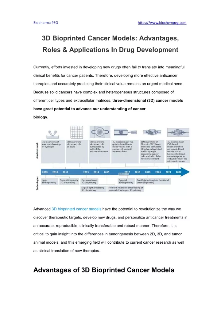
Biopharma PEG https://www.biochempeg.com 3D Bioprinted Cancer Models: Advantages, Roles & Applications In Drug Development Currently, efforts invested in developing new drugs often fail to translate into meaningful clinical benefits for cancer patients. Therefore, developing more effective anticancer therapies and accurately predicting their clinical value remains an urgent medical need. Because solid cancers have complex and heterogeneous structures composed of different cell types and extracellular matrices, three-dimensional (3D) cancer models have great potential to advance our understanding of cancer biology. Advanced 3D bioprinted cancer models have the potential to revolutionize the way we discover therapeutic targets, develop new drugs, and personalize anticancer treatments in an accurate, reproducible, clinically transferable and robust manner. Therefore, it is critical to gain insight into the differences in tumorigenesis between 2D, 3D, and tumor animal models, and this emerging field will contribute to current cancer research as well as clinical translation of new therapies. Advantages of 3D Bioprinted Cancer Models
Biopharma PEG https://www.biochempeg.com An ideal 3D bioprinted cancer model can precisely reproduce the in vivo environment of a specific tumor, including its perfusion vessels. This enables multiple biochemical assessments of tumor cell behavior that mimic the in vivo environment. Indeed, gene expression analysis showed that compared with 2D cultures, 3D bioprinted cancer models could show immunoglobulin production, expression of proinflammatory molecules, activation of cytokines and/or chemokines, upregulation of cell-cell adhesion pathways, and reduction of proteins associated with cell division and DNA replication. These differences provide insights into how the 3D environment affects cancer cell growth, migration, invasion, stem cells, and gene expression. In addition, the elasticity, plasticity, and mechanical properties of the original tumor ECM can be modeled by using specific matrix materials. For example, hepatogenic decellularized ECM and mammary decellularized ECM have retained microstructures and
Biopharma PEG https://www.biochempeg.com ultrastructures that, together with growth factors bound and sequestered in the matrix, control cell location and orientation. The decellularized ECM scaffolds printed by digital light processing (DLP) technology enable accurate spatial cell deposition, thereby preserving these tissue-specific growth factors. DLP-based models have also been used to study the initial stages of pancreatic ductal adenocarcinoma development. Blood vessels play a crucial role in tumor proliferation, oxygen diffusion, angiogenesis, endovascular and extravasation. Therefore, the realization of functional vascular networks in biomimetic tumor models is essential to maintain cell viability and reveal the close relationship between tumor and blood vessels. This dynamic environment can be studied by 3D bioprinted cancer models containing vasculature, including how circulating cancer cells interact with stromal cells and infiltrating immune cells, the exchange of secreted factors between different cell types, the response to external stimuli, and the behavioral adaptation of cancer cells to the metastatic microenvironment. Each cancer has a unique TME that includes various healthy functional cell types, such as stromal cells, vascular cells, and immune cells. However, non-bioprinted 3D cancer models, which are implemented by means of structures such as hydrogel constructs, polymer scaffolds, microcarrier beads, and hanging droplets, do not allow spatiotemporal control of tissue formation and do not allow long-term observation of dynamic changes. 3D bioprinted models can overcome these limitations by reconstructing the entire TME, including its functional and structural hierarchies, thus faithfully mimicking the complex in vivo tumor tissue structure at high resolution and maintaining the viability and function of patient-derived tissue. The Important Role of 3D Bioprinted Cancer Models Simulating a Metastatic Niche
Biopharma PEG https://www.biochempeg.com An important challenge in cancer research is to construct in vitro models that can reproduce natural metastatic niches. In addition to differences in ECM properties between metastatic sites, the interaction between invading cancer cells and the TME within the metastatic niche is critical in mediating the metastatic cascade. In a 3D bioprinted model simulating the bone metastatic niche, MDA-MB-231 breast cancer cells were co-cultured with osteoblasts and human bone marrow mesenchymal stem cells (MSCs) to mimic the bone TME. The proliferation rate of MSCs and osteoblasts decreased within 5 days after the addition of cancer cells, suggesting that breast cancer cells induce osteolysis in tumor bone. In addition, breast cancer cells in this model showed increased secretion of the proangiogenic factor VEGF and decreased alkaline phosphatase activity, which are markers of new bone formation. Simulating tumor blood vessel Improved understanding of tumor cell-endothelial cell interactions could reveal important mechanisms of tumor metastasis and angiogenesis. By 3D bioprinting, breast cancer microspheres were generated, which encapsulated microfibers containing human umbilical vein endothelial cells (HUVEC). When co-cultured with breast cancer cells, HUVEC elongates toward cancer cells outside the fibers, which remain exclusively within the fibers and form vascular-like cavities within the fibers. This finding shows the potential of co-cultured 3D bioprinted cancer models to reshape the interaction between cancer cells and endothelial cells. Anti-tumor immunity Bringing immune cells from the TME and periphery into 3D bioprinted models can provide a reproducible platform to study human anticancer immune responses, thereby generating tumor models suitable for understanding tumor biology and drug testing. For example, in a 3D bioprinted model consisting of bladder cancer cells, fibroblasts, HUVEC,
Biopharma PEG https://www.biochempeg.com and monocytes, treatment with Bacille Calmette-Guerin (BCG) resulted in increased monocyte proinflammatory cytokine secretion and decreased cancer cell growth. Currently, several 3D bioprinted models have been developed to rapidly and reliably evaluate the efficacy of immune modulators and cell-based cancer immunotherapies. For example, 3D bioprinted models have been used to evaluate chimeric antigen receptor (CAR) T-cell therapy for neuroblastoma. Brain malignancies face considerable therapeutic challenges, in part because of their unique brain TMEs that promote tumor progression. A DLP-based 3D bioprinted glioblastoma model has been developed that mimics the brain TME and contains glioma stem cells, astrocytes, neural precursor cells, and macrophages. The model is also capable of analyzing macrophage phenotypes and detecting multiple transcriptional changes that occur as a result of cancer cells interacting with the TME. In this model, macrophages recruited by cancer cells acquire a glioma-associated phenotype that promotes tumorigenesis. By combining different technologies and tuning the desired tissue-like properties and cellular components, 3D bioprinted models can serve as valuable tools for studying TME and cancer immunology. Drug Development Applications of 3D Bioprinted Cancer Models 3D bioprinting enables the assembly of cells and ECMs to form 3D constructs that demonstrate the complexity of cancer tissues and serve as a robust and reproducible platform for the discovery of new therapeutic targets, preclinical testing of anticancer drugs, and the development of personalized cancer therapies.
Biopharma PEG https://www.biochempeg.com Drug Efficacy Evaluation 3D-printed biological cancer models have been used in the screening and discovery of a variety of drugs. ECM properties, such as density and composition, influence drug spread and tumor penetration, and some 3D bioprinted tumor models take these factors into account. In an iterative 3D bioprinting approach using GP-118 patient-derived gastric adenocarcinoma cells suspended in a gelatin-alginate-matrix biomaterial, this 3D bioprinted gastric adenocarcinoma model is chemo-resistant to Docetaxel, 5-fluorouracil, and cisplatin and can be used to assess resistance to developing drugs. 3D bioprinted models have also been used to evaluate the therapeutic effects of monoclonal antibodies. For example, metuzumab, an anti-CD174 antibody used to treat a variety of cancers, researchers used heat-sensitive biomaterials to 3D bioprint microfluidics composed of SMMC-7721 liver cancer cells and HUVECs. Higher doses of metuzumab were required to inhibit cancer cell migration and proliferation in 3D models
Biopharma PEG https://www.biochempeg.com compared with 2D cultures. Additionally, the incorporation of human peripheral blood mononuclear cells into the 3D bioprinted model allowed the investigators to assess metuzumab-induced ADCC cytotoxicity, an important aspect of therapeutic antibodies. Drug screening platform In addition to evaluating tumor response to drugs, 3D bioprinting platforms can help high-throughput screening of compounds and approval of drugs for different diseases or indications. Whole exome sequencing (WES) can identify the mutation profiles of patient cancer samples and predict drug sensitivity by associating these profiles with specific drugs that target mutations. Target Discovery The addition of perfusable vascular systems to multicellular 3D bioprinted models may further improve drug screening platforms. For example, a 3D bioprinted microengineered glioblastoma model is being developed that contains perfusable capillaries lined with endothelial and pericytes and connected to a peripheral blood pump. This 3D model can reflect the heterogeneity of glioblastoma samples, and the tumor cells in this 3D model are transcriptionally more similar to in vivo glioblastoma tumor cells than 2D cultures derived from the same cells. Notably, this model shows upregulation of P-selectin, whereas glioblastoma cells grown in 2D medium do not express P-selectin and are not affected by P-selectin inhibitors, suggesting that the use of 3D bioprinted cancer models may reveal therapeutic targets that cannot be detected by conventional 2D culture. In conclusion, 3D bioprinted cancer models have been shown to better reflect tumor heterogeneity, TME complexity, cancer cell behavior, gene expression signatures, and drug response than traditional 2D culture methods. These models also provide a platform for studying parameters of cancer therapeutic approaches that cannot be adequately studied using conventional 2D culture methods or simple 3D models.
Biopharma PEG Clinical Trials https://www.biochempeg.com Various ongoing clinical trials are evaluating the predictive power of 3D cancer models for drug screening, target discovery, and personalized therapy. For example, an ongoing clinical trial is evaluating 3D bioprinted liver cancer models to predict the response of chemotherapy to colorectal cancer as well as liver metastases from colorectal cancer (NCT04755907). Another trial uses 3D bioprinting of hyaluronic acid-gelatin biomaterials to create organ-like models of myeloma (NCT03890614) and aims to create patient-specific bioimprinted models to study myeloma biology and chemotherapy sensitivity. These clinical studies provide proof of concept for the feasibility of using 3D bioprinted cancer models to accurately model patient tumors and their dynamic microenvironment and predict treatment outcomes. Conclusion 3D bioprinted cancer models have the potential to transform the way we study, diagnose, prevent and treat cancer. The commercialization of these models, especially in drug development and testing, is expected to yield substantial economic benefits. In addition, advanced 3D bioprinting technologies, combined with machine learning and AI-based omics approaches, may uncover fundamental mechanisms of cancer biology, reveal novel biomarkers and drug targets, and advance the development of effective personalized cancer treatments. develop. As a leading PEG supplier, Biopharma PEG specializes exclusively in the development and manufacturing of high-quality PEG products and derivatives. We supply AC-PEG-AC, AC-PEG-RGD and 8-ArmPEG-AC which can used in 3D bioprinting. PEG-based hydrogels are most used polymers in the 3D printing techniques due to their good biocompatibility in both in vitro and in vivo conditions. We can also supply 4-ArmPEG-SG, 4-ArmPEG-SS, 8-ArmPEG-SG and 8-ArmPEG-SS which can be
Biopharma PEG https://www.biochempeg.com used to crosslink into degradable PEG hydrogels. Reference: [1]. 3D bioprinted cancer models: from basic biology to drug development. Nat Rev Cancer.2022 Oct 24. Related articles: [1]. Strategies Of Oral Drug Delivery: From Prodrug, Nanoparticles to 3D Printing [2]. Polyethylene Glycol (PEG) Hydrogel Based 3D Bioprinting [3]. A Covalently Crosslinked Bioink Used In Three-dimensional Cell Cultures of 3D Bioprinting [4]. The Role of PEGylated Materials In 3D Bioprinting
