Download
1 / 23
320 likes | 816 Views
Antioxidant Enzymes. Maria Holmstrom Qiang Zhang Nicole Milkovic Erin Rosenbaugh. Introduction to Antioxidant Enzymes. Beal, Nature. 2006 Oct 19;443(7113):787-95. Superoxide dismutases. Catalyzes the dismutation of superoxide into oxygen and hydrogen peroxide Diffusion limited

E N D
Antioxidant Enzymes Maria Holmstrom Qiang Zhang Nicole Milkovic Erin Rosenbaugh
Introduction to Antioxidant Enzymes Beal, Nature. 2006 Oct 19;443(7113):787-95.
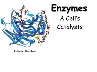
Superoxide dismutases • Catalyzes the dismutation of superoxide into oxygen and hydrogen peroxide • Diffusion limited • Found in nearly all oxygen-exposed cells • Categorized by metal prosthetic group • Cu/Zn, Mn, Fe or Ni
Species • B. Subtilis MnSOD (sodA) • E. Coli MnSOD (sodA) and FeSOD (sodB) • S. Cerevisiae CuZnSOD (sod1) and MnSOD (sod2) • H. Sapiens CuZnSOD (sod1), MnSOD (sod2) and EC-SOD (CuZn, sod3)
Localization • Bacteria • SOD A cytoplasm • SOD B cytoplasm • Eukaryotes • SOD1 cytosol • SOD2 mitochondrial matrix • SOD3 (humans) glycated and secreted into the extracellular space, and subsequently anchored to heparan sulfate proteoglycans
Catalytic site, bovine SOD1 Image from: Pelmenschikov & Siegbahn, Inorg. Chem, 2005
M(n+1)+ − SOD + O2− → Mn+ − SOD + O2 Mn+ − SOD + O2− + 2H+ → M(n+1)+ − SOD + H2O2 Image from: Pelmenschikov & Siegbahn, Inorg. Chem, 2005
Catalase Catalase is a common enzyme found in nearly all living organisms that are exposed to oxygen, where it functions to catalyze the decomposition of hydrogen peroxide to water and oxygen First noticed by Louis Jacques Thénard in 1818 First named as catalase by Oscar Loew in 1900 Catalase is a tetramer highest turnover numbers
Cofactors • Heme • Manganese
Distribution among organisms • All known animals use catalase in every organ, with particularly high concentrations occurring in the liver • Catalase is also universal among plants, and many fungi are also high producers of the enzyme • Catalase has also been observed in some anaerobic microorganisms
Reconstructed phylogenetic tree of 70 typical catalases from all main living kingdoms ANTIOXIDANTS & REDOX SIGNALING Volume 10, Number 9, 2008
Catalase genes • Bacillus subtilis: katA(vegetative catalase 1), katX(catalase in spores), katE(catalase 2) • E. Coli: katE(HPII(III)), katG(HPI), katP(EHEC-catalase) • S. Cerevisiae: CTA1(Catalase A), CTT1(Ctt1p ) • H. Sapiens: CAT(Catalase)
Location • Intracellular • Extracellular • Cell surface • Periplasm • Cytoplasm • Cytosol • Glyoxysome • Mitochondrion
Introduction to Peroxiredoxin • Widely distributed thiol-based group of enzymes that catalyze the reduction of H2O2, organic hydroperoxides (ROOH), and peroxynitrite • ROOH +2e- ROH + H2O • 3 Classes: Typical 2-Cys, Atypical 2-Cys, 1-Cys
Isoforms of Mammalian Peroxiredoxins Wood ZA et al. (2003) Structure, mechanism and regulation of peroxiredoxins. TRENDS Bio Sci 28:32-40
Peroxiredoxin Mechanism Wood ZA et al. (2003) Structure, mechanism and regulation of peroxiredoxins. TRENDS Bio Sci 28:32-40
1.65Å Structure Of Prx From Aeropyrumpernix K1 Complexed With H2O2 Nakamura Tet al. (2010) Crystal structure of peroxiredoxin from Aeropyrumpernix K1 complexed with its substrate, hydrogen peroxide. J. Biochem. 147:109-115
Glutathione (GSH) and the Glutathione Peroxidase (GPx) Activity of an Erythrocyte Factor Protect Hemoglobin from Oxidative Breakdown A B Crystalline hemogloblin + AA + GSH + NaN3 Erythrocyte hemosylate containing hemogloblin Crystalline hemogloblin Boiled Enzyme A. Effect of azide (catalase inhibitor) and GSH (reduced glutathione) on the coupled oxidation of hemoglobin by ascorbic acid (AA) B. Concentration-dependent effects of erythrocyte enzyme preparation on choleglobin formation Journal of Biochemistry. Gordon C. Mills, 1957
Function of Glutathione Peroxidase (GPx) GPx4 ROOR' (lipid hydroperoxidase) + electron donor (2 e-) + 2H+ ROH + R'OH • GPx and GSH remove intracellular hydrogen peroxide and hydroperoxides to protect cellular components from oxidative damage/modifications • GPX reduces many reactive oxygen species (e.g., lipid hydroperoxides (ROOR’) to alcohols and to reduce free hydrogen peroxide to water) • Glutathione system often functions in parallel with thioredoxin system to regulate the redox homeostasis in cells GPx 2 GSH (reduced glutathione) + H2O2 GSSG (oxidized glutathione) + 2 H2O
Mechanism of Glutathione Peroxidase Mechanism for GPx catalytic cycle Active Site of Gpx Selenocysteine residue Reaction 3 is rate limiting step (1) Peroxide(e.g., H202) reduction and oxidation of the selenolate anion/ selenol (E-Se- orE-Se-H) to selenenic acid (E-SeOH) (2)Selenenic acid reacts with GSH to produce seleno-sulfide adduct (E-Se-SG) (3) 2nd GSH molecule attacks E-Se-SG to regenerate active GPx and GSSG GPx 2 GSH (reduced glutathione) + H2O2 GSSG (oxidized glutathione) + 2 H2O Prabhakar, R. et al. Biochemistry, 2005
Subcellular Localization of GPx in Mammalian Cells Extracellular fluids: GPx3 cGPX or GPX1- cytosol giGPX or GPX2- cytosol, vesicular structures (external cell surface?) Glycosylated GPX, pGPX or GPX3- extracellular, compartments (e.g., plasma) PHGPX or GPX4- mitochondrial membranes, nucleus, nucleolus, cytosol GPx4 GPx4 GPx1, Gpx2, GPx4 Modified from geneticssuite.net/node/11
Species and Tissues that express Glutathione Peroxidase GPx gene clusters from Group I → metazoans (animal kingdom) Group II → fungi, proteobacteria, cyanobacteria, algae Group III → plant kingdom GPX1- found in nearly all tissues GPX2- gastrointestinal tract GPX3- extracelluar fluids and low levels in plasma; mRNA predominately in kidney GPX4- ubiquitously expressed; membrane fractions of testis Margis R. et al. FEBS Journal. 2008;
Antioxidant Enzymes Beal, Nature. 2006 Oct 19;443(7113):787-95.