Download
1 / 78
850 likes | 1.93k Views
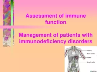
Assessment and Management of Patients with Endocrine Disorders. By Linda Self. Glands of the Endocrine System. Hypothalamus Posterior Pituitary Anterior Pituitary Thyroid Parathyroids Adrenals Pancreatic islets Ovaries and testes. Hypothalamus. Releasing and inhibiting hormones

E N D
Assessment and Management of Patients with Endocrine Disorders By Linda Self
Glands of the Endocrine System • Hypothalamus • Posterior Pituitary • Anterior Pituitary • Thyroid • Parathyroids • Adrenals • Pancreatic islets • Ovaries and testes
Hypothalamus • Releasing and inhibiting hormones • Corticotropin-releasing hormone • Thyrotropin-releasing hormone • Growth hormone-releasing hormone • Gonadotropin-releasing hormone • Somatostatin-=-inhibits GH and TSH
Anterior Pituitary • Growth Hormone-- • Adrenocorticotropic hormone • Thyroid stimulating hormone • Follicle stimulating hormone—ovary in female, sperm in males • Luteinizing hormone—corpus luteum in females, secretion of testosterone in males • Prolactin—prepares female breasts for lactation
Posterior Pituitary • Antidiuretic Hormone • Oxytocin—contraction of uterus, milk ejection from breasts
Adrenal Cortex • Mineralocorticoid—aldosterone. Affects sodium absorption, loss of potassium by kidney • Glucocorticoids—cortisol. Affects metabolism, regulates blood sugar levels, affects growth, anti-inflammatory action, decreases effects of stress • Adrenal androgens—dehydroepiandrosterone and androstenedione. Converted to testosterone in the periphery.
Adrenal Medulla • Epinephrine and norepinephrine serve as neurotransmitters for sympathetic system
Thyroid • Follicular cells—excretion of triiodothyronine (T3) and thyroxine (T4)—Increase BMR, increase bone and calcium turnover, increase response to catecholamines, need for fetal G&D • Thyroid C cells—calcitonin. Lowers blood calcium and phosphate levels
Parathyroid • Parathyroid hormone—regulates serum calcium
Pancreatic Islet cells • Insulin • Glucagon—stimulates glycogenolysis and glyconeogenesis • Somatostatin—decreases intestinal absorption of glucose
Kidney • 1, 25 dihydroxyvitamin D—stimulates calcium absorption from the intestine • Renin—activates the RAAS • Erythropoietin—Increases red blood cell production
Ovaries • Estrogen • Progesterone—inportant in menstrual cycle,*maintains pregnancy,
Testes • Androgens, testosterone—secondary sexual characteristics, sperm production
Thymus • Releases thymosin and thymopoietin • Affects maturation of T lymphocetes
Pineal • Melatonin • Affects sleep, fertility and aging
Prostaglandins • Work locally • Released by plasma cells • Affect fertility, blood clotting, body temperature
Assessment • Health history—energy level, hand and foot size changes, headaches, urinary changes, heat and cold intolerance, changes in sexual characteristics, personality changes, others • Physical assessment—appearance including hair distribution, fat distribution, quality of skin, appearance of eyes, size of feet and hands, peripheral edema, facial puffiness, vital signs
Diagnostic Evaluation • Serum levels of hormones • Detection of antibodies against certain hormones • Urinary tests to measure by-products (norepinephrine, metanephrines, dopamine) • Stimulation tests—determine how an endocrine gland responds to stimulating hormone. If the hormone responds, then the problem lies w/hypothalmus or pituitary • Suppression tests—tests negative feedback systems that control secretion of hormones from the hypothalamus or pituitary.
Disorders of the Pituitary Pituitary Tumors • Eosinophilic tumors may result in gigantism or in acromegaly. May suffer from severe headaches, visual disturbances, decalcification of the bone, endocrine disturbances • Basophilic tumors may cause Cushing’s syndrome w/features of hyperadrenalism, truncal obesity, amenorrhea, osteoporosis • Chromophobic tumors—90% of pituitary tumors. Present with lowered BMR, obesity, somnolence, scant hair, low body temp, headaches, visual changes
Growth hormone deficiency in childhood will result in primary dwarfism.
Pituitary Tumors—Assessment and Diagnostic Findings • H&P • Vision tests • CT, MRI • Serum levels of pituitary hormones, others
Diabetes Insipidus • Deficiency of ADH • Excessive thirst, large volumes of dilute urine • Can occur secondary to brain tumors, head trauma, infections of the CNS, and surgical ablation or radiation • Nephrogenic DI—relates to failure of the renal tubules to respond to ADH. Can be related to hypokalemia, hypercalcemia and to medications (lithium demeocycline)
Manifestations • Excessive thirst • Urinary sp. gr. of 1.001.1.005
Assessment and Diagnostic Findings • Fluid deprivation test—withhold fluids for 8-12 hours. Weigh patient frequently. Inability to slow down the urinary output and fail to concentrate urine are diagnostic. Stop test if patient is tachycardic or hypotensive • Trial of desmopressin and IV hypertonic saline • Monitor serum and urine osmolality and ADH levels
Pharmacologic Tx and Nursing Management • DDAVP—intranasal bid • Can be given IM if necessary. Every 24-96h. Can cause lipodystrophy. • Can also use Diabenese and thiazide diuretics in mild disease as they potentiate the action of ADH • If renal in origin—thiazide diuretics, NSAIDs (prostaglandin inhibition) and salt depletion may help • Educate patient about actions of medications, how to administer meds, wear medic alert bracelet
SIADH • Excessive ADH secretion • Retain fluids and develop a dilutionalhyponatremia • Often non-endocrine in origin—such as bronchogenic carcinoma • Causes: Disorders of the CNS like head injury, brain surgery, tumors, infections or medications like vincristine, phenothiazines, TCAs or thiazide diuretics • Meds can either affect the pituitary or increase sensitivity to renal tubules to ADH • Management: eliminate cause, give diuretics (Lasix), fluid restriction, I&O, daily wt., lab chemistries
SIADH • Restoration of electrolytes must be gradual • May use 3% NaCl in conjunction with Lasix
Thyroid • T3 and T4 • Need iodine for synthesis of hormones—excess will result in adaptive decline in utilization called the Wolf-Chaikoff mechanism • Thyroid is controlled by TSH • Cellular metabolism, brain development, normal growth, affect every organ in the body • T3 is five times as potent as T4 • Calcitonin—secreted in response to high levels of serum calcium, increases deposition in the bone
Thyroid • Inspect gland • Observe for goiter • Check TSH, serum T3 and T4 • T3 resin uptake test useful in evaluating thyroid hormone levels in patients who have received diagnostic or therapeutic dose of iodine. Estrogens, Dilantin, Tagamet, Heparin, amiodarone, PTU,steroids and Lithium can cloud the accuracy • T3 more accurate indicator of hyperthyroidism according to text
Thyroid • Antibodies seen in Hashimoto’s, Grave’s and other auto-immune problems. • Radioactive iodine uptake test measures rate of iodine uptake. Patients with hyperthyroidism exhibit a high uptake, hypothyroidism will have low uptake • Thyroid scan—helps determine the location, size, shape and size of gland. “Hot” areas (increased function) and “cold” areas (decreased function) can assist in diagnosis.
Nursing Implications • Be aware of meds patient is taking (see list in text) that can affect accuracy of testing • Also be aware if patient is taking multivitamins and food supplements
Hypothyroidism • Most common cause is Hashimoto’s thyroiditis • Common in those previously treated for hyperthyroidism • Atrophy of gland with aging • Medications like lithium, iodine compounds, antithyroid meds can cause • Radiation treatments to head and neck • Infiltrative diseases like amyloidosis, scleroderma • Iodine deficiency and excess • Hypothalamic or pituitary abnormality • More common in women, especially over age 50
Manifestations • From mild symptoms to myxedema • Myxedema –accumulation of mucopolysaccharides in sc and interstitial tissues. Is the extreme form of hypothyroidism. Can progress to shock. • S/S—fatigue, hair loss, dry skin, brittle nails, numbness and tingling of the fingers, amenorrhea, weight gain, decreased heart rate and temperature, lassitude, cognitive changes, elevated cholesterol levels, constipation, hypotension
Pharmacologic Management of hypothyroidism • Levothyroxine is preferred agent • Dosage is based on TSH • Desiccated thyroid used infrequently due to inconsistent dosing • Angina can occur when thyroid replacement is initiated as it enhances effects of cardiovascular catecholamines (in pt. w/pre-existent CAD). Start at low dose. • Hypnotics and sedatives may have profound effects on sensorium
Management in Myxedema • Cautious fluid replacement • Glucose to restore to normal glycemic levels • Avoid rapid overheating due to increased oxygen demands but keep warm • May give levothyroxine intravenously With recovery, • Modify activity • High fiber foods • Home health for follow-up
Hyperthyroidism • Extreme form is Grave’s disease • Caused by thyroiditis, excessive amount thyroid hormone, abnormal output by immunoglobulins • Is more common in women
Manifestations of hyperthyroidism • Thyrotoxicosis—nervousness, irritable, apprehensive, palpitations, heat intolerance, skin flushing, tremors, possibly exophthalmos • Have an increased sensitivity to catecholamines • Can occur after irradiation or presence of a tumor
Assessment and Diagnosis • Thyroid thrill and or bruit may be present • Thyroid may be enlarged • Decreased TSH, increased free T4 and an increased radioactive iodine uptake
Management • Reduce thyroid hyperactivity—usually use radioactive iodine, antithyroid meds or surgery) • Beta blockers • Can be relapse with antithyroid meds
Pharmacologic Therapy • Irradiation with administration of radioisotope iodine 131—initially may cause an acute release of thyroid hormones. Should monitor for thyroid storm • S/S of thyroid storm—high fever. Tachycardia, delirium, chest pain, dyspnea, palpitations, weight loss, diarrhea, abdominal pain • Management of thyroid storm—oxygen, IV fluids with dextrose, hypothermic measures, steroids to treat shock or adrenal deficiency, iodine to decrease output of T4, beta blockers, PTU or Tapazole impedes formation of thyroid hormone and blocks conversion of T4 to T3
Antithyroid Medications • PTU—propylthiouracil—blocks synthesis of hormones • Tapazole (methimazole)—blocks synthesis of hormones. More toxic than PTU. • Sodium Iodide-suppresses release of thyroid hormone • SSKI (saturated solution of potassium chloride)– suppresses release of hormones and decreases vascularity of thyroid. Can stain teeth • Dexamethazone—suppresses release of thyroid hormones
Surgical Management • Reserved for special circumstances, e.g. large goiters, those who cannot take antithyroid meds, or who need rapid normalization • Subtotal thyroidectomy • Before surgery, give PTU until s/s of hyperthyroidism have disappeared • Iodine may be used to decrease vascularity
Nursing Management • Reassurance r/t the emotional reactions experienced • May need eye care if has exophthalmos • Maintain normal body temperature • Adequate caloric intake • Managing potential complications such as dysrhythmias and tachycardias • Educate about potential s/s of hypothyroidism following any antithyroid tx.
Parathyroid Glands • Parathormone maintains sufficient serum calcium levels • Excess calcium can bind with phosphate and precipitate in various organs, can cause pancreatitis • Hyperparathyroidism will cause bone decalcification and development of renal calculi • More common in women • Secondary hyperparathyroidism occurs in those with chronic renal failure and renal rickets secondary to excess phosphorus retention (and increased parathormone secretion)
Manifestations of Hyperparathyroidism • May be asymptomatic • Apathy, fatigue, muscle weakness, nausea, vomiting, constipation, hypertension and cardiac dysrhythmias • Excess calcium in the brain can lead to psychoses • Renal lithiasis can lead to renal damage and even failure • Demineralization of bones with back and joint pain, pain on weight bearing, pathologic fractures • Peptic ulcers and pancreatitis can also occur
Assessment and Diagnostic Findings • Persistent elevated calcium levels • Elevated serum parathormone level • Bone studies will reveal decreased density • Double antibody parathyroid hormone test is used to distinguish between primary hyperparathyroidism and malignancy • Ultrasound, MRI, thallium scan, fine needle biopsy also can be used to localize cysts, adenomas, or hyperplasia
Management • Recommended treatment for hyperparathyroidism is surgical removal • Hydration therapy necessary to prevent renal calculi • Avoid thiazide diuretics as they decrease renal excretion of calcium • Increase mobility to promote bone retention of calcium • Avoid restricted or excess calcium in the diet • Fluids, prune juice and stool softeners to prevent constipation • Watch for s/s of tetanypostsurgically(numbness, tingling, carpopedal spasms) as well as cardiac dysrhythmias and hypotension
Hypercalcemic crisis • Seen with levels greater than 15mg/dL • Can result in life-threatening neurologic, cardiovascular and renal symptoms • Treatments include: hydration, loop diuretics to promote excretion of calcium, phosphate therapy to promote calcium deposition in bone and reducing GI absorption of calcium • Give calcitonin or mithramycin to decrease serum calcium levels quickly
Hypoparathyroidism • Seen most often following removal of thyroid gland, parathyroid glands or following radical neck surgery • Deficiency of parathormone results in increased bone phosphate and decreased blood calcium levels • In absence of parathormone, there is decreased intestinal absorption of dietary calcium and decreased resorption of calcium from bone and through kidney tubules
Clinical Manifestations of Hypoparathyroidism • Irritability of neuromuscular system • Tetany—hypertonic muscle contractions , numbnes, tingling, cramps in extremities, laryngeal spasm, bronchospasm, carpopedal spasm ( flexion of the elbows and wrists, dorsiflexion of the feet), seizures