Download
1 / 61
720 likes | 1.3k Views
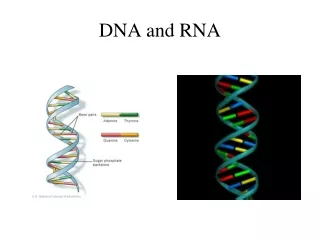
DNA and RNA. Dr. Sugandhika Suresh Department of Biochemistry. Features of the DNA double helix. 2 DNA strands per molecule Right-handed helix 2 chains are antiparallel Sugars and phosphates –outside Bases inside -- “stacked like pennies” Bases are bonded together by H-bonds

E N D
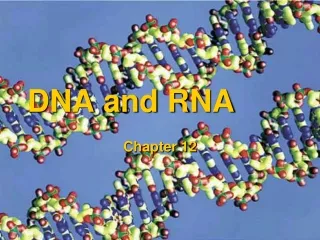
DNA and RNA Dr. Sugandhika Suresh Department of Biochemistry
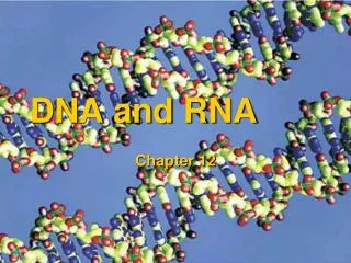
Features of the DNA double helix • 2 DNA strands per molecule • Right-handed helix • 2 chains are antiparallel • Sugars and phosphates –outside • Bases inside -- “stacked like pennies” • Bases are bonded together by H-bonds • Specific base pairings are observed - complementary • A pairs with T • C pairs with G • 10 base pairs per turn • Spacing causes a major and a minor groove
Two strands are twisted together around • a common axis • Right handed
Right-Handed vs.Left-Handed Helices • The helix is right-handed • As it spirals away from you, the helix turns in a clockwise direction
The two strands are antiparallel • One runs in the 5’ to 3’ direction and the other 3’ to 5’ • Deoxyribose phosphate backbone -hydrophilic
DNA Can Form Alternative Types of Double Helices • The DNA double helix can form different types of secondary structure • The predominant form found in living cells is B-DNA • However, under certain in vitro conditions, A-DNA and Z-DNA double helices can form
DNA forms B-form A-form Z-form
A-DNA • Right-handed helix • 11 bp per turn • Occurs under conditions of low humidity • Little evidence to suggest that it is biologically important • Z-DNA • Left-handed helix • 12 bp per turn • Its formation is favored by • GG-rich sequences, at high salt concentrations • Cytosine methylation, at low salt concentrations • Evidence from yeast suggests that it may play a role in transcription and recombination
Denaturation of DNA • Denaturation by heating. • How is it observed? • A260 • For dsDNA, A260=1.0 for 50 µg/ml • For ssDNA and RNA A260=1.0 for 38 µg/ml • For ssoligonuleotides A260=1.0 for 33 µg/ml • Hyperchromic shift The T at which ½ the DNA sample is denatured is called the melting temperature (Tm)
The two strands of the double helix separate reversibly at high temperatures The temperature at which this “denaturation” or “melting” occurs depends on the pH and salt concentration, and increases with the GC content of the DNA. (The curves drawn here are schematic.) If the temperature is lowered, the strands recombine. The rate of reassociation is inversely proportional to the complexity of the DNA.
dA nucleotides dG ssDNA dU dsDNA dC Double-stranded and single-stranded DNA differ in their optical absorption at 260 nm The absorbance of double-stranded DNA (dsDNA) at 260 nm is less than that of either single-stranded DNA (ssDNA) or the free bases. This is called “hypochromism.” The conjugated p-electron systems of the purine & pyrimidine bases absorb strongly in the UV. (That’s why UV light is mutagenic and carcinogenic.)
Importance of Tm • Critical importance in any technique that relies on complementary base pairing • Designing PCR primers • Southern blots • Northern blots • Colony hybridization
Factors Affecting Tm • G-C content of sample • Presence of intercalating agents (anything that disrupts H-bonds or base stacking) • Salt concentration • pH • Length
Renaturation • Strands can be induced to renature (anneal) under proper conditions. Factors to consider: • Temperature • Salt concentration • DNA concentration • Time
T G A Histone octamer C GC TA Histone proteins GC CG TA AT AT CG 2 nm GC TA Packaging DNA B DNA Helix
Histone proteins 2 nm Packaging DNA T G A Histone octamer C GC TA GC CG TA AT AT CG B DNA Helix GC TA
11 nm Histone proteins Nucleosome 2 nm Packaging DNA T G A Histone octamer C GC TA GC CG TA AT AT CG B DNA Helix GC TA
DNA-histone octamer H1 Links Nucleosomes together
Nucleosomes: +H1 -H1
GC CG TA AT AT CG GC TA Packaging DNA
Packaging DNA GC CG TA AT AT CG GC TA
Packaging DNA 11 nm GC CG TA AT 30 nm 200 nm AT CG GC TA Protein scaffold “Beads on a string” Looped Domains Tight helical fibre
Packaging DNA 11 nm Nucleosomes 30 nm 700 nm 200 nm T Looped Domains Tight helical fibre G C A 2 nm Protein scaffold B DNA Helix Metaphase Chromosome
A packaged chromosome Chromatid Identical chromatid Replication Anaphase Chromosomes, Chromatids and Centromeres Chromosome arm Two identical chromosomes Centromere Chromosome arm
RNA structure and function Objectives • The differences between DNA and RNA • The structure and function of RNAs
RNA & DNA: Similarities Both RNA & DNA: • Unbranched polymers • Polynucleotides • Contain phosphodiester bonds
RNA & DNA: Differences • RNA • Single-Strand (mostly) • Cytoplasm (mainly) • AGCU • Modified bases • Ribose • Protein Biosynthesis • Post-transcriptional events • DNA • Double • Nucleus • d AGCT • Deoxyribose • Storage &transfer • DNA Repair
Biological roles of RNA • RNA is the genetic material of some viruses • RNA functions as the intermediate (mRNA) between the gene and the protein-synthesizing machinery. • RNA functions as an adaptor (tRNA) between the codons in the mRNA and amino acids.
4.Through sequence complementarity, RNA serves as a regulatory molecule to bind to and interfere with the translation of certain mRNAs; or as a recognition molecule to guide many post-transcriptional processing steps. 5.Through the tertiary structures, some RNAs function as enzymes to catalyze essential reactions in the cell (RNase P ribozyme, large rRNA in ribosomes, self-splicing introns, etc).
RNA Structure • The primary structure of an RNA strand is much like that of a DNA strand • RNA strands are typically several hundred to several thousand nucleotides in length • In RNA synthesis, only one of the two strands of DNA is used as a template
Components unique to RNA Replaces Deoxyribose Replaces Thymine
RNA Primary Structure (-e) Structure of RNA backbone 5' (-e) (-e) (-e) 3' • RNA chain directionality: 5'3' • Backbone carries charge (-e) on each nucleotide • Formation of an RNA structure requires cations
Although usually single-stranded, RNA molecules can form short double-stranded regions • This secondary structure is due to complementary base-pairing • A to U and C to G • This allows short regions to form a double helix • RNA double helices typically • Are right-handed • Have the A form with 11 to 12 base pairs per turn • Different types of RNA secondary structures are possible
Structures of RNA • Primary structure • 2.Sequence complementarity: base pairing as DNA • 3.Secondary structure • 4. Tertiary structure
Primary structure RNA STRUCTURE RNA contains ribose and uracil and is usually single-stranded
U 2.Sequence complementarity: inter- and intra-molecular base pairing RNA STRUCTURE (1) Watson-Crick base pairing G-C A-U
RNA chains fold back on themselves to form local regions of double helix similar to A-form DNA RNA STRUCTURE (2) 2nd structure elements hairpin RNA helix are the base-paired segments between short stretches of complementary sequences, which adopt one of the various stem-loop structures bulge loop
Complementary regions Noncomplementary regions Held together by hydrogen bonds Also called hair-pin Have bases projecting away from double stranded regions
The double helical structure of RNA resembles the A-form structure of DNA. • The minor groove is wide and shallow, but offers little sequence-specific information. • The major groove is so narrow and deep that it is not very accessible to amino acid side chains from interacting proteins.