Download
1 / 61
660 likes | 1.21k Views
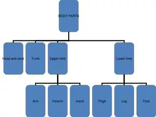
Pelvis & Lower limb. X-Ray of the pelvis and lower limb. Joint of lower limb. Hip # - types. Femoral head usually the result of high energy trauma and a dislocation of the hip joint often accompanies this fracture.

E N D
Pelvis & Lower limb X-Ray of the pelvis and lower limb
Hip # - types • Femoral head usually the result of high energy trauma and a dislocation of the hip joint often accompanies this fracture. • Femoral neck subcapital, or intracapsulardenotes a # adjacent to the femoral head in the neck between the head and the greater trochanter • Intertrochanteric a break in which the # line is between the greater and lesser trochanter on the intertrochanteric line . • Subtrochanteric # actually involves the shaft of the femur immediately below the lesser trochanter, may extend down the shaft of the femur.
MRI PELVIS Multiple pelvic fractures
X-Ray of pelvic fractures A suspected pelvic fracture requires the following plain x-rays (1)Standard AP view (2)Outlet view (3)Inlet view These may be supplemented by CT scans for further clarification and evaluation and for patient management. Assessment for associated soft tissue injuries such as urethral and/or bladder injuries is a must especially in unstable pelvic fractures
AP View of the pelvis: the X-ray beam is perpendicular to the pelvis and the film cassette
Inlet view of the pelvis: The X-ray beam is directed to the mid pelvis at 60 degree angle to the film cassette.
Outlet view of the pelvis: The X-ray beam is directed from the feet to the symphysis at 40 degree angle to the film cassette.
Stable pelvic fracture • Plain film of the pelvis shows the normal arcuate line of the right SI joint and fracture of the left superior pubic rami. • CT shows fracture of the anterior sacrum near the left SI joint
Unstable pelvic fracturepelvic ring interrupted • Plain x-ray AP view showing bilateral separation of SI joints and wide diastasis of symphysis pubis
Acetabular fractures • Acetabular fractures occur primarily in young adults as a result of high energy trauma. • The contact area between femoral head & acetabulum will be decreased. • Mal union of the acetabular fracture will lead to post traumatic arthritis. • 2 basic x-ray views are required for assessment of acetabular fracture:- (1) AP view followed by- (2) 45 degree internal oblique view ( Judet view)
Dislocation of the hip joint Types: Posterior dislocation • Most common type of dislocation. • Femoral head is lateral & superior to acetabulum • Posterior rim of acetabulum is usually fractured • Associated sciatic nerve injury in 10% Anterior dislocation • Femoral head displaced into the obturator, pubic or iliac region Internal dislocation • Always associated with acetabular fracture • Femoral head protrudes into the pelvic cavity.
Dislocation of HipAnterior dislocation of hip.Plain x-ray showing abduction and external rotation.
Posterior dislocation of hipPlain x-ray showing adduction & internal rotation of the hip
Fractures of the proximal femur Classification Intracapsular fractures involving femoral head or neck: • Capital: uncommon • Subcapital: common • Transcervical: uncommon Extracapsular fractures involving the trochanters • Intertrochanteric • Subtrochanteric
Fracture proximal femurIntertrochanteric fracture (Extacapsular fracture)
(A) Undisplaced intertrochanteric fracture not clearly seen on plain x-ray.(B) Corresponding MR clearly displays the fracture
Avascular necrosis of femoral head ( post fracture complication)
Fractures of distal femur Classification 1-Supracondylar • Non displaced • Displaced • Impacted • Comminuted 2-Condylar 3-Intercondylar
Fracture of the distal femurComminuted intercondylar fracture
Transmission of energy resulting in concomittant injury patternsIn all orthopaedic injuries it is mandatory to rule out additional injuries to the joint above and below to rule out ipsilateral fractures of the femur, acatabulum and patella epeciallyafter high energy vehicle trauma
Standard x-ray views for the knee(A) AP view (B) Lateral view (C) Tunnel view (D) Skyline view
X-ray lateral view of the knee showing joint effusion following trauma: displacement of the black translucent fat line in supra patella region
Tibial plateau fractureAP view: Vertical lucent fracture line ( black arrows) with cortical step (white arrow) Cross table lateral view: fat fluid level in supra patella region
X-ray lateral view of the ankle showing effusion: displacement of the translucent fat line (arrows)
Lower limbMassoneuve fracture : disruption of the tibiofibular syndesmoses
(A) Bimalleolar fracture : Horizontal fracture line in medial malleolus and oblique fracture line in lateral malleollus (B) Trimalleolar fracture: Above plus fracture of posterior malleolus.