Download
1 / 66
660 likes | 1.05k Views
STROKE, TIA, AND OTHER CENTRAL FOCAL CONDITIONS. Dee Mortensen PGY2 November 10, 2005 Tintinalli Ch. 228. Background. Third leading cause of death in US Leading cause of adult disability 700,000 patients/year 1/3 of patients younger than 65. Definition of Stroke.

E N D
STROKE, TIA, AND OTHER CENTRAL FOCAL CONDITIONS Dee Mortensen PGY2 November 10, 2005 Tintinalli Ch. 228
Background • Third leading cause of death in US • Leading cause of adult disability • 700,000 patients/year • 1/3 of patients younger than 65
Definition of Stroke • Any disease process that disrupts blood flow to a focal region of the brain.
Stroke Types • 80% ischemic • Thrombosis • Embolism • Hypoperfusion • 20% hemorrhagic • Intracerebral • Subarachnoid
Ischemic Strokes • Thrombosis-most common cause • Etiology • Atherosclerotic disease-most common • Vasculitis • Dissection • Polycythemia • Hypercoagulable states • Infectious Diseases-HIV, TB, syphilis
Ischemic Strokes • 1/5th due to Embolism • Etiology • Cardiac • Valvular Vegetations • Mural thrombi- caused by A-fib, MI, or dysrhythmias • Paradoxical emboli-from ASD, VSD • Cardiac tumors-myxoma • Fat emboli • Particulate emboli – IV drug injections • Septic Emboli
Ischemic Strokes • Hypoperfusion- less common mechanism • Typically caused by cardiac failure • More diffuse injury pattern vs thrombosis or embolism • Usually occur in watershed regions of brain
Hemorrhagic Strokes • Intracerebral hemorrhage (ICH) - approx. 10% of all strokes • Risk Factors • HTN • Increasing Age • Race: Asians and Blacks • Amyloidosis- esp. in the elderly • AVMs or tumors • Anticoagulants/Thrombolitic use • History of previous stroke • Tobacco, ETOH, and cocaine use
Hemorrhagic Stroke • Subarachnoid hemorrhage (SAH) • Result from rupture of berry aneurysm or rupture of AVMs
Cerebral Anatomy • Vascular circulation: Anterior and Posterior • Anterior circulation • Origin: carotid system • supplies 80% brain- optic nerve, retina, frontoparietal and anterotemporal lobes of brain
Anterior Circulation Anatomy • Common carotid artery • Divides into Internal and External carotids at angle of mandible • Internal carotid artery – terminates at anterior and middle cerebral artery at the circle of Willis • Ophthalmic artery – 1st branch off internal carotid -supplies optic nerve and retina
Posterior Circulation Anatomy • Posterior circulation: supplies 20% of brain • Derived from vertebral arteries • Posterior circulation supplies brainstem, cerebellum, thalamus, auditory centers and visual cortex
Ischemic Pathophysiology • Neurons are very sensitive to cerebral blood flow and die within minutes of complete cessation • Extent of injury depends on vessel involved and presence or absence of collateral blood flow • Penumbra • Reversibly injured neurons surrounding the primary injury with collateral circulation, which can be preserved with proper timely intervention
Hemorrhagic Pathophysiology • In ICH and SAH, intracranial pressure rises following vascular rupture with resulting global hypoperfusion • Marked reduction in perfusion occurs near the hematoma as a result of local compression
Clinical Features • Stroke presentation often subtle and varied • Key aspects in determining the underlying cause and location of the lesion include: • History • Physical Exam • Neurologic Exam
History • History of: • HTN • CAD • DM • Previous TIA in same vascular distribution • Symptomatic deficits that wax and wan • Gradual onset • Suggests: atherosclerotic disease and thrombosis
History • History of • A-Fib • Valvular replacement • Recent MI • Multiple TIAs involving different vascular distributions • Sudden onset of symptoms • Suggests: Embolism
History • History of : • Recent neck injury-MVA, Sports injury • Chiropractic manipulation • Suggests: Carotid dissection
History • History of: • Straining or coughing immediately preceding symptoms • Suggests: ruptured aneurysm
History • History of: • Sudden onset of symptoms • Headache (minority of patients with ischemic stroke) • Suggests: Hemorrhagic stroke
Physical Exam • Not inclusive, but some pointers: • Signs of emboli- Janeway lesions, Osler nodes • Bleeding dyscrasia- ecchymosis, petechiae • Papilledema- mass lesion, HTN crisis, cerebral vein thrombosis • Carotid bruit or murmurs- vascular or cardiac etiol.
Neurologic Exam (see Ch 226) • National Institutes of Health (NIH) Stroke Scale- correlates to infarct volume • Six major areas: • LOC • Visual Assessment • Motor Function • Cerebellar Function • Sensation and Neglect • Cranial Nerves
Stroke Syndromes • Classic physical exam findings that assist in localizing the lesion.
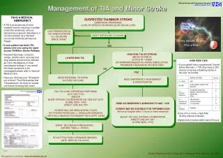
Ischemic Stroke Syndrome • Transient Ischemic Attack (TIA) • Neurologic deficit that resolves within 24 hours • Most TIAs resolve < 30 minutes • Approx. 10% of patients will have a stroke in 90 days • Half of these in just 2 days
Ischemic Stroke Syndromes • Anterior Cerebral Artery Infarction • Contralateral weakness/numbness greater in leg than arm • Dyspraxia • Speech perseveration • Slow responses
Ischemic Stroke Syndromes • Middle cerebral artery occlusion • Dominant Hemisphere (usually the left) • Contralateral weakness/numbness in arm and face greater than leg • Contralateral hemianopsia • Gaze preference toward side of infarct • Aphasia (Wernicke’s -receptive, Broca’s -expressive or may have both) • Dysarthria
Ischemic Stroke Syndromes • Middle cerebral artery occlusion • Nondominant hemisphere • Contralateral weakness/numbness in arm and face greater than in the leg • Constructional Apraxia • Dysarthria • Inattention, neglect, or extinction
Ischemic Stroke Syndromes • Posterior Cerebral Artery Infarct • Often unrecognized by patient- minimal motor involvement • Light-touch/pinprick may be significantly reduced • Visual cortex abnormalities also minimal
Ischemic Stroke Syndromes • Vertebrobasilar Syndrome • Posterior circulation supplies brainstem, cerebellum, and visual cortex • Dizziness, vertigo, diplopia, dysphagia, ataxia, cranial nerve palsies, and b/l limb weakness, singly or in combination • HALLMARK: Crossed neurological deficits: ipsilateral CN deficits with contralateral motor weakness
Ischemic Stroke Syndromes • Lateral Medullary (Wallenburg) Syndrome • Specific post. Circulation infarct involving vertebrobasilar and/or post inferior cerebellar Art. • Signs: • Ipsilateral loss of facial pain and temperature with contralateral loss of these senses over the body • Gait and limb ataxia • Partial ipsilateral loss of CN V, IX, X, and XI • Ipsilateral Horner Syndrome may be present
Ischemic Stroke Syndromes • Basilar Artery Occlusion • Severe quadriplegia • Coma • Locked-in syndrome-complete muscle paralysis except for upward gaze
Ischemic Stroke Syndromes • Cerebellar Infarction-subset of post. circ. infarcts • Symptoms: “drop attack” with sudden inability to walk or stand, often a/w vertigo, HA, nausea/vomiting, neck pain • Diagnosis: MRI, MRA as bone artifact obscures CT • Cerebral edema develops w/in 6-12 hrs → increased brainstem pressure and decreased LOC • Treatment: decrease ICP and emergent surgical decompression
Ischemic Stroke Syndrome • Lacunar Infarction • Infarction of small penetrating arteries in pons and basal ganglia • Associated with chronic HTN present in 80-90% • Pure motor or sensory deficits • Arterial Dissection • Often a/w severe trauma, headache, and neck pain hours to days prior to onset of neuro symptoms • HTN risk factor for spontaneous dissection
Hemorrhagic Syndromes • Intracerebral Hemorrhage • ICH – sudden onset HA, N/V, elevated BP • Progressive focal neurologic deficits over minutes • Patients may rapidly deteriorate • Exertion commonly triggers symptoms • Bleeding localized to putamen, thalamus, pons-pinpoint pupils, and cerebellum
Hemorrhagic Syndromes • Cerebellar Hemorrhage • Sudden onset dizziness, vomiting, truncal ataxia, inability to walk • Possible gaze palsies and increasing stupor • Treatment: urgent surgical decompression or hematoma evacuation
Hemorrhagic Syndrome • Subarachnoid hemorrhage • Severe HA, vomiting, decreasing LOC • HA- often occipital or nuchal in location • Sudden onset of symptoms– history may reveal activities a/w ↑ HTN such as defecation, coughing or intercourse
Diagnosis-Critical Pathway • History • Last moment patient known to be normal • Initial orders • ECG, Cardiac Enzymes, CBC, Coags, Type/Screen, Lytes, glucose, Renal function studies, +/- drug screen, Noncontrast CT-head • Review alteplase inclusion/exclusion criteria
Diagnostic Tests • Emergent noncontrast CT of head • Differentiate hemorrhage vs ischemia • MOST ischemic strokes (-) by CT for at least 6 hrs • Hypodensity indicating infarct seen 24-48 hrs • Can identify hemorrhage greater than 1cm, and 95% of SAH • If CT (-) but still considering SAH may do L.P.
Diagnostic Tests • Depending on circumstances, other helpful tests • Echocardiogram – identifies mural thrombus, tumor, valvular vegetations in suspected cardioembolic stroke • Carotid duplex -for known/suspected high grade stenosis • Angiography – “gold standard” identifies occlusion or stenosis of large and small vessels of head/neck, dissections and aneurysms • MRI scan – identifies posterior circulation strokes better and ischemic strokes earlier than CT • Emergent MRI- considered for suspected brainstem lesion or dural sinus thrombosis • MRA scan – identifies large vessel occlusions – may replace angiography in the future
Differential Diagnosis • Ddx of Acute Stroke (not inclusive) • Epidural/subdural hematoma • Hyponatremia • Brain tumor/abscess • Postictal paralysis (Todd paralysis) • Hypertensive encephalopathy • Meningitis/encephalitis • Hyperosmotic coma
Differential Diagnosis Cont. • Wernicke Encephalopathy • Drug toxicity (lithium, phenytoin, carbamazepine) • Complicated Migraine • Bells palsy • Multiple sclerosis • Meniere’s disease • Labyrinthitis
Special Populations In Stroke • Sickle Cell Disease (SCD) • Most common cause of ischemic stroke in children • 10% of patients with Sickle Cell Disease have stroke by age 20 • SCD-↑ frequency of cerebral aneurysm—think SAH • Treatment: emergent simple or exchange transfusion to decrease HbS to < 30%, thus improving blood flow and oxygen delivery to infarct zone
Special Populations In Stroke • Young Adults (age 15 to 50) • 20% of ischemic strokes due to arterial dissection • Often preceded by minor trauma • Cardioembolic etiologies- MVP, rheumatic heart disease, or paradoxical embolism • Migrainous stroke- infarction a/w typical attack • Air embolism-scuba diving or recent invasive procedure • Drugs: heroin, cocaine, amphetamines
Special Populations In Stroke • Pregnancy • ↑risk during peripartum and up to 6 weeks postpartum • Contributors to risk-preeclampsia/eclampsia, decrease in blood vol. and hormonal status following birth
Ischemic Stroke Management • General Management • A, B, Cs • IV, oxygen, monitor, elevate head of bed slightly • E.D. protocols/Notify stroke team • Treat dehydration and hypotension • Avoid overhydration – cerebral edema • Avoid IVF with glucose – except if hypoglycemic • Fever – worsens neurologic deficits
Ischemic Stroke Management • Hypertension • Treatment indicated for SBP > 220 mm Hg or mean arterial pressure > 130 mm Hg • Lowering BP too much reduces perfusion to penumbra converting reversible injury to infarction • Use easily titratable Rx (labetalol or enalaprilat) • SL Ca-channel blockers should be avoided
Management of HTN cont. • Thrombolytic candidates- use NTG paste or Labetalol to reduce BP < 185/115 to allow tx • Requirements for more aggressive treatment exclude the use of tissue plasminogen activator.
Thrombolysis Background • NIH/NINDS study • 624 patients, RDBPC trial IV tPA vs placebo • Treatment w/in 3 hrs of onset • At 3 months pts tx’d with tPA were at least 30% more likely to have minimal/no disability…absolute favorable outcome in 11-13 percent • 6.4% of patients treated with tPA developed symptomatic ICH compared with 0.6% in placebo group • Mortality rate at 3 months not significantly different • tPA group had significantly less disability • FDA approved in 1996
tPA Dose and Complications • IV tPA –Total dose 0.9 mg/kg, max. 90mg • 10% as bolus, remaining infusion over 60 min. • BP and Neuro checks q 15 min x 2 hrs initially • Treatment must begin w/in 3 hrs of symptoms and meet inclusion and exclusion criteria • No ASA or heparin given x 24 hrs after tx
Emergent Mngt of HTN during/following rtPA in Acute Stroke • Monitor BP closely • q 15 min x 2 hrs, then q 30 min x 6 hrs, then q 60 min for 24 hr Total • If SBP 180-230 or DBP 105-120 mmHg • 10 mg labetalol IVP q 10-20 min, max 150 mg • If SBP > 230 or DBP 121-140 mmHg • 10 mg labetalol may repeat q 10-20 min, max 150 mg • If BP not controlled by labetalol then consider nitroprusside (0.5-1.0mcg/kg/min), continuous arterial monitoring advised • If DBP > 140 mmHg • Infuse sodium nitroprusside (0.5-1.0mcg/kg/min), continuous arterial monitoring advised