Download
1 / 27
270 likes | 486 Views
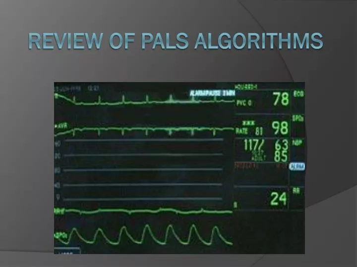
Review of PALS Algorithms. Algorithms. Bradycardia with a Pulse Stable Cardiopulmonary status Cardiopulmonary Compromise Tachycardia with Pulses and Poor Perfusion Sinus Tachycardia Supraventricular Tachycardia Ventricular Tachycardia Pulseless Arrest

E N D
Algorithms • Bradycardia with a Pulse • Stable Cardiopulmonary status • Cardiopulmonary Compromise • Tachycardia with Pulses and Poor Perfusion • Sinus Tachycardia • Supraventricular Tachycardia • Ventricular Tachycardia • Pulseless Arrest • Ventricular Fibrillation vs. Ventricular Tachycardia • Asystole vs. PEA • Septic Shock
Steps involved when called to evaluate a patient • Help can be called for at any time!!! The sooner the better! • #1 • LOOK AT YOUR PATIENT!! • Sick or not sick • #2 • Examine • #3 • Cardiac Monitor • #4 • Check wires/tubing • #5 • Call for help and Establish Roles • #6 • Identify Algorithm • #7 • Resuscitation Supplies/Meds
Sick or Not Sick • http://www.youtube.com/watch?v=XJ-ON24aO9s&feature=BFa&list=PLF7E5E6EAB1933606
Examine • Get history (RN/family concerns, HPI, hospital course) • Vital signs (trends and current) • Physical Exam (ABC’s) • Airway • Breathing • Cardiovascular
Physical Exam • Airway • Can patient speak or cry? • Look: for respiratory distress (i.e. grunting, flaring retractions), choking, cyanosis • Listen: air movement in neck and chest, quality (stridor, wheeze, etc..), I:E, RR • Feel: movement from mouth, nose, chest rise, crepitus • Assess: can airway be maintained with basic maneuvers/positioning, suction
Physical Exam • Breathing • Is patient moving sufficient air in and out to maintain effective oxygenation and ventilation? • Look: RR, trachea position, symmetry of chest rise, accessory muscles, skin color • Listen: symmetry and quality, adequacy of air movement, intrathoracic sounds (stridor, crackles, wheeze) • Feel: subcutaneous air, tenderness, instability of chest wall • Assess: stable or not? Respiratory failure? BMV? Intubate? Trumpets?
Physical Exam • Cardiovascular • Adequate circulation to support end-organ function? • Look: • poor perfusion: cyanosis, mottling, pallor, altered mental status • Chest trauma: asymmetry of chest expansion • Jugular Venous Distention • Listen • Heart rate: tachycardicvs. bradycardicvs. normal • Heart tones: murmur, rub, gallop • Breath sounds: ralesvs. wheeze • Diminished Breath sounds • Feel • Central pulses, temperature of skin, capillary refill • Assess • Adequate vs. inadequate vs. absent
Cardiac Monitor • Know what the numbers on the monitor mean • Make sure connections are correct and wires/leads actually attached to the patient • Lead placement: White is right, Smoke (black) over fire (red) • Waveform • Heart rate from leads and pulse oximeter should correlate • If patient on oxygen, make sure connected appropriately to wall with no kinks and cannula/mask placed appropriately
Establish Roles • Code Team • Team Leader • Airway Physician • Float Physician • Medication Nurse • Bedside Nurse • Circulating Nurse • Documenter • Assistants
What makes a Rhythm Shockable? • The heart is active, but in a life-threatening and dysfunctional pattern. In Ventricular Tachycardia, the heart is unable to pump blood effectively as it is beating too quickly to fill. This will ultimately lead to ventricular fibrillation. At this point the electrical activity in the heart becomes chaotic – again preventing the heart from pumping effectively. Over time, the fibrillation will decrease and the heart will become asystolicfrom lack of appropriate oxygenation.
What makes a Rhythm Shockable? • Defibrillation – for UNORGANIZED rhythms • Therapeutic delivery of an unsynchronized electrical current through the myocardium over a brief period to terminate the dysrhythmia • Does not jump start the heart • Purpose is to depolarize the ventricular cells simultaneously (including fibrillating cells) asystole natural pacemakers will resume normal activity
What makes a Rhythm Shockable? • Synchronized Cardioversion – for ORGANIZED • Delivery of a shock to the heart to terminate a rapid dysrhythmia that is times to avoid vulnerable periods in the cardiac cycle (peak to end of T wave) • Heart cells will contract simultaneously interrupting and terminating the abnormal electrical rhythm without damaging the heart allowing the sinus node to resume normal pacemaker activity