Download
1 / 23
1.11k likes | 3.69k Views
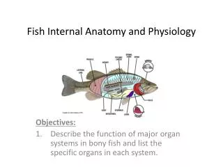
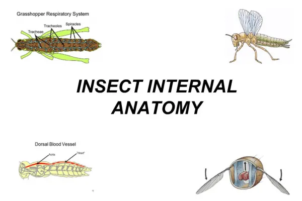
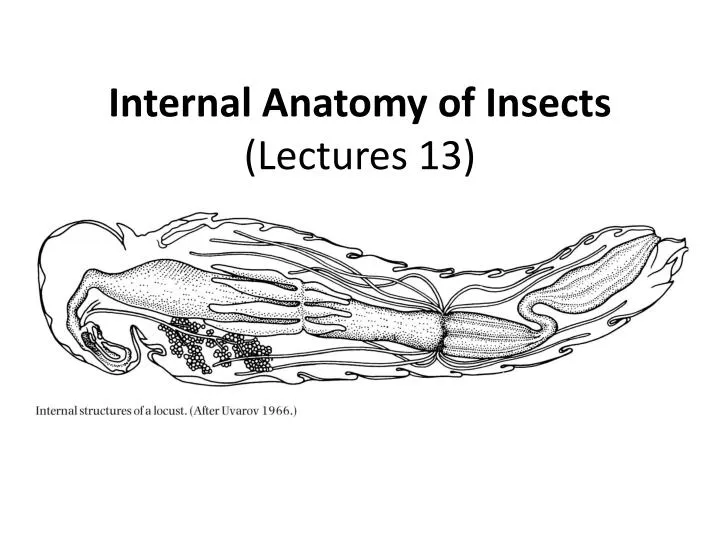
Internal Anatomy of Insects (Lectures 13). What’s inside the bug?. Internal anatomy. Muscular system – muscles and apodemes Nervous system – neurons, CNS Endocrine system – hormones, endocrine cells Circulatory system – hemolymph , vessels Respiratory system – spiracle, trachea

E N D
Internal anatomy • Muscular system – muscles and apodemes • Nervous system – neurons, CNS • Endocrine system – hormones, endocrine cells • Circulatory system – hemolymph, vessels • Respiratory system – spiracle, trachea • Digestive system – gut, fat body • Excretory system – Malpighian tubules, rectum • Reproductive system – male and female structures
Muscular system • All insect muscles are striated • Hierarchical organization: muscle fiber, myofibril, thin filament (actin), thick filament (myosin) • Sarcomere: basic contractile unit of muscle • Basically same as vertebrate muscles
Muscle attachments • Insect muscles must attach to the inner surface of an internal skeleton • Muscles need to be fused with exoskeleton by growing tonofibrillae • Muscle attachment sites: apodemes (internal ridges), apophyses (elongated arms)
How insects perceive the world chemical, tactile visual visual chemical chemical chemical, tactile acoustic tactile
Nervous system • Neuron, axon, dendrite, synapse • Basically same as vertebrates
Central Nervous System (CNS) • Ganglia – nerve centers consisting of cell bodies of interneurons and motor neurons aggregated with fibers interconnecting all types of nerve cells • Primitively a pair of ganglia per body segment • Fusing and reduction of ganglia
Central Nervous System (CNS) • Ganglia – nerve centers consisting of cell bodies of interneurons and motor neurons aggregated with fibers interconnecting all types of nerve cells • Primitively a pair of ganglia per body segment • Fusing and reduction of ganglia
Insect brain • Protocerebrum – optic lobes • Deutocerebrum – antennae • Tritocerebrum – signals from body • Suboesophageal ganglion – mouthparts
Insect brain optic lobe protocerebrum antennal nerve deutocerebrum frontal ganglion tritocerebrum suboesophageal ganglion Lateral Frontal
Insect brain optic lobe protocerebrum antennal nerve deutocerebrum frontal ganglion tritocerebrum suboesophageal ganglion Lateral Frontal
Endocrine System • Hormones – signal chemicals that travel through circulatory system • Endocrine cells – produce hormones • Neurosecretory cells: modified neurons • Corpora cardiaca: store and release neurohormones • Prothoracic glands: secrete ecdysone (molting hormone) • Corpora allata: secrete juvenile hormone
Rhodniussp. Vector of Chagas disease One of Wigglesworth’s experiments in determining the hormonal actions in insects V.B. Wigglesworth, Father of Insect Endocrinology
Types of insect hormones • Ecdysteroid: steroid that promotes molting activity • Ecdysone and 20-hydroxyecdysone • Juvenile hormone (JH): control of metamorphosis, regulation of reproductive development • JH-I, JH-II, JH-III, etc. • Neurohormone: aka neuropeptides, regulate development, homeostasis, metabolism, and reproduction • Several hundreds identified
Mechanism of phase transformation: An overview Favorable environmental conditions leading to concentration Locusts detect change in density through cephalic and thoracic pathways Release of serotonin leading to behavioral gregarization Prolonged high density leading to synthesis and release of a neuropeptide, [His7]-corazonin, or dark-color-inducing neuropeptide from CC Collective movement achieved by locusts’ aligning to moving objects, reinforced by cannibalism
Prolonged high density leads to synthesis and release of a neuropeptide, [His7]-corazonin Isolated nymph Crowded nymph Isolated nymph crowded from 3rdinstar Isolated nymph injected with [His7]-corazonin Tawfik et al. 1998 (PNAS)
Circulatory System • Open circulatory system with the blood (hemolymph) occupying the general body cavity, known as a hemocoel • Hemolymph flows by muscular contractions and contractions of a dorsal vessel • Hemocoel is divided into three major sections: dorsal pericardinal sinus, perivisceral sinus, and ventral perineural sinus
Hemolymph circulation • Main pump: dorsal vessel (anterior: aorta, posterior: heart) • Ostia: segmentally arranged openings • Dorsal and ventral diaphragm, formed of connective tissue and segmental pairs of alary muscles • Appendage circulation unidirectional powered by accessory pulsatile organs
Dorsal vessel Alary muscles
Dorsal vessel Alary muscles
Hemolymph • < 20% of body weight • Colorless (typically), yellow, green, blue, red • Ions (chloride [most abundant], sodium, potassium, calcium, magnesium, etc) • Free amino acids • Proteins • Storage proteins (hexamerins) • Lipid transport proteins (lipophorins) • Binding proteins (ferritins, JH-binding proteins) • Carbohydrates (trehalose, glucose) • Protection and defense (injury, immune response, chemical deterrence)