Download
1 / 33
400 likes | 673 Views
Silicon Photomultipliers and their bio-medical applications. E.Grigoriev 3,4 , A.Akindinov 4 , M.Breitenmoser 3 , S.Buono 1 , E.Charbon 2 , I.Desforges 3 , C.Niclass 2 , R.Rocca 1 1 AAA (Advanced Accelerator Applications), Saint Genis, France

E N D
Silicon Photomultipliers andtheir bio-medical applications E.Grigoriev3,4, A.Akindinov4, M.Breitenmoser3, S.Buono1, E.Charbon2, I.Desforges3, C.Niclass2, R.Rocca1 1 AAA (Advanced Accelerator Applications), Saint Genis, France 2 EPFL, AQUA group, Lausanne, Switzerland 3 FORIMTECH S.A., Geneva, Switzerland 4 ITEP, Moscow, Russia E.Grigoriev – EuroMedim 2006
Outline • Introduction: from silicon SPAD to SiPM and SPAD arrays • SPAD arrays – features and applications • SiPM – current status and main features • SiPM – examples of applications in physics • SiPM – examples of applications in medicine • Prospects of use in bio-medical applications • Summary E.Grigoriev – EuroMedim 2006
p+ n-well n+ n-well p-substrate SPAD – principle of operation • SPAD – Single Photon Avalanche Diode • Operates at Vop=Vbd+DV when a self-sustaining avalanche process can be initiated by a single charge-carrier [Haitz, 1965] E.Grigoriev – EuroMedim 2006
photon arrival Vop V SPAD recharge Vbd Avalanche quenching SPAD: main characteristics • Typical size 10-100 mm • Vbreakdown 20-40 V • QE peak ~ 45 % at ~500 nm • Cjunction ~ 10-100 fF • Dark count rate ~ 0.1-10 Hz / mm2 • Q=C(Vop-Vbd)=M (“gain”) Rochas, Ph.D. Thesis, EPFL, 2003 E.Grigoriev – EuroMedim 2006
Vop VDD R T q q V Vp+ IA IA Passive and Active Recharge VDD digital pulse OUT Vp+ = VDD + |VH| > Vbd trecharge < 40 nsec trecharge > 100 nsec Niclass and Charbon, ISSCC 2005 E.Grigoriev – EuroMedim 2006
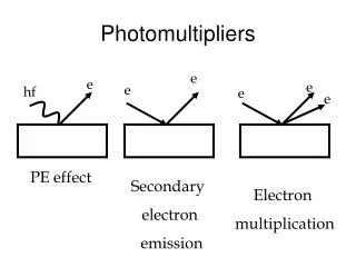
SPAD limitations Capabilities of SPAD quite limited: • Small geometrical acceptance • 1 photon at a time + ~40 nsec for recharge NB: many fluorescent processes are characterized by lifetimes in 0.1-10 nanoseconds band time-resolved measurement impossible with one SPAD Need for devices with many sensitive elements and/or high dynamic range E.Grigoriev – EuroMedim 2006
Two types of SPAD arrays SiPM • Dense packing (fill factor ~ 50-80%) • Common readout • Special optical isolation (MESA technology,…) • Dynamic range ~ Npix • Passive recharge (trec > 100 nsec) SPAD chips • Loose packing (fill factor ~ 1-6%) • Individual addressing • No special optical isolation (standard CMOS process) • Dynamic range = 1 • Active recharge (trec< 40 nsec) E.Grigoriev – EuroMedim 2006
Guard Ring Osc. LFSR 1k pixel array R O W D E C O D E R Anode Logic Gates 2.8mm COL. DECODER 2.5mm CMOS 32x32 Pixel Array • 0.8 mm CMOS • Active area diam. 7 mm • Pitch = 58 mm • Fill factor ~ 1% • QE at 635 nm ~ 12% • Time jitter ~ 50 psec • Dead time < 40 nsec Niclass,Charbon et al., JSSC 05 E.Grigoriev – EuroMedim 2006
Example: 3D Vision • Lateral resolution: • 64x64 pixels • Depth resolution: • 1.3mm • Range: • 3.75m [Niclass and Charbon, ISSCC 2005] E.Grigoriev – EuroMedim 2006
CMOS Deep-submicron SPAD array • Features • World’s first DSM SPAD array • World’s smallest pitch (25mm) and largest single photon streak camera (112x4 pixels) • Event-driven readout • Fill factor: 6% [Niclass, Sergio, Charbon, DATE 06] E.Grigoriev – EuroMedim 2006
Readout -Voper SiPM design principles • Maximize geometrical fill factor • Common readout of all channels (analog sum of quasi-digital signals) • Passive quenching with individual resistors in each pixel • Good optical isolation between pixels (MESA,…) G. Bondarenko, V. Golovin, M. Tarasov, patent N 2142175 (Russia 1999) E.Grigoriev – EuroMedim 2006
SiPM layout N. Basharuli, G. Bondarenko, V. Golovin et al. , Como 2001 E.Grigoriev – EuroMedim 2006
SiPM pulseheight spectra Spectrum from a pulsed low-light source. Peak widths reflect spread in pixel capacitance values and breakdown voltages A.Akindinov et al., Beaune 2005 to be published in NIM E.Grigoriev – EuroMedim 2006
SiPM noise spectrum (log scale) 1e 1000 2e 100 3e 10 1 SiPM optical cross-talk Probability to have more than one pixel fired from a single primary e-h pair = (N n+1)/ (N n ) A. Akindinov, V. Golovin, E. Grigoriev et al., Proceedings of 8th ICATPP conference Villa Erba, Como, 2004 E.Grigoriev – EuroMedim 2006
SiPM photon detection efficiency P.D.E. = Q.E. * g* R εg – geometrical fill-factor R – probability to initiate Geiger avalanche Q.E. – quantum efficiency Y. Musienko, S. Reucroft, J. Swain Northeastern University, Boston This spectral sensitivity well matches the wavelenghs of most of fluorescent proteins A.V. Akindinov et al., Instrum. Exp. Tech. 48-3 (2005) 355 E.Grigoriev – EuroMedim 2006
SiPM timing properties Single photoelectron timing resolution for SiPM and PMT B. Dolgoshein et al. / Nucl. Instr. Methods A 504 (2003) 48–52 Resolution for multiphoton events improves as 1/sqrt(N) E.Grigoriev – EuroMedim 2006
SiPM characteristics • operating voltage ~ 20-40 V • power consumption ~ 50 W / mm2 • single-photon response ~ 105-106e • optical cross-talk ~ 10% • peak detection efficiency ~ 25% at 520nm • timing resolution ~ 100 psec • typical size ~ 1 mm2 • dynamic range ~ 1000 • low sensitivity to ionizing particles • non-sensitivity to magnetic field • RT operation, low temperature dependence • mechanical and electrical robustness • low price E.Grigoriev – EuroMedim 2006
SiPM features • Large photosensitive area • Large dynamic range • Deadtime negligible if Nph << Npixels • Possibility of dark count discrimination (dark count is predominantly single-electron) BUT: • Significant dark count rate (~105-106 Hz / mm2) • Enhanced optical cross-talk (~10%) Therefore area is practically limited to few mm2 Best application - in time-correlated measurements with fibers, microlenses or in microdevices E.Grigoriev – EuroMedim 2006
Physics: particle detectors with SiPM Cosmic triggering with scintillating tiles and WLS fibers (START) Particle tracking with Scintillating fibers – Beam Profile Monitor 2 planes of 80 tiles Z. Sadygov et al. / Nucl. Instr. Methods A 550 (2005) 212–216 • A.Akindinov, E. Grigoriev, V. Golovin et al., • - Nucl. Instrum. Methods A539 (2005) 172-176 E.Grigoriev – EuroMedim 2006
Physics: calorimetry with SiPM Hadron calorimeter (CALICE Collab.) B. Dolgoshein, M. Danilov et al. / Nucl. Instr. Methods A 540 (2005) 368–380 E.Grigoriev – EuroMedim 2006
Medicine: SciFi+SiPM probe for diagnostics and radio-guided surgery AAA/FORIMTECH design (patent pending) In collaboration with University Hospitals of Grenoble, Geneva and Lausanne Work supported by INTERREG Grant 49/BL/9.3/3 E.Grigoriev – EuroMedim 2006
SciFi probe Principle of directional selectivity Extremely high sensitivity to positrons has been already demonstrated! Numerous applications in local dynamic multi-positional detection of different radio-tracers (intravascular, transcutaneous, intracavital) AAA / FORIMTECH design (patent pending) Work supported by INTERREG Grant 49/BL/9.3/3 E.Grigoriev – EuroMedim 2006
SciFi probe: directional resolution versus threshold for different fiber diameters E.Grigoriev – EuroMedim 2006
WLS-optical fibers Matrix of scintillating elements or single plate SiPM matrix New ideas & applications NB SiPM can be easily transformed in a multichannel device by modifying the top Al layer Large Area Radiation Imager • Applications: • PET • Portal imaging with plastic scintillator and high threshold • Imaging of large objects with high- energy g, n, p FORIMTECH design (patent pending) E.Grigoriev – EuroMedim 2006
New ideas & applications Universal time-resolved fluorescence probe. Fiber(s) can be clear or WLS, can be inserted in a catheter or seringe needle for study of dynamic processes using optically stimulated fluorescence Simulteneous detection of several fluorochromic labels FORIMTECH design (patent pending) E.Grigoriev – EuroMedim 2006
Optical Dielectrophoresis Monitoring New ideas & applications Single photon detection islands Artist rendering of a dielectrophoresis system based on [Romani et al, ISSCC 04] with the proposed addition of optical monitoring E.Grigoriev – EuroMedim 2006 [Romani et al. ISSCC 04]
Confocal optics Confocal optics Detector Laser Timing Analysis (Two-Photon) Fluorescence Lifetime Imaging (FLIM) • Simplified FLIM optical setup (Calcium-sensitive dyes) • Transmission configuration Advantage of SPAD array: • Can process multiple points at a time through deep sub-micron highly parallel sensor • picosecond accuracy with non-cooled sensor [Agronskaia et al., 2004] E.Grigoriev – EuroMedim 2006
New ideas (medicine, biology, environment protection) Single-photon sensitivity, RT operation, good timing resolution allow SiPM and SPAD arrays to be used in compact and robust devices which can measure molecular signatures and are based on time-resolved fluorescence, delayed luminescence, photon scattering or radio-tracing: • DNA analysis, • measuring protein dynamics using light scattering, • two-photon fluorescence microscopy • automated DNA sequencing machines • particle and droplet sizing • optical biopsy • study of cancer processes using fluorescent proteins • fluorescence or diffuse optical tomography • implantable or endovascular probes this list can be very long… Potential partners interested to develop jointly any of these are welcome! E.Grigoriev – EuroMedim 2006
Recently approved project: RAPSODI (FP6 / CRAFT) GOAL: implementation of SiPM in a range of medical devices for dosimetry and radio-protection Participants: • University of Insubria (Como) – coordinator • ITEP (Moscow) • University of Technology (Krakow) • FORIMTECH (Geneva) • PTW (Freiburg) • SensL (Cork) • Jiry-Plch SMM (Prague) E.Grigoriev – EuroMedim 2006
SUMMARY • SiPMs and SPAD arrays have already demonstrated superior performances in particle physics and several other applications • Their commercial availability and customisation open numerous wide opportunities for replacement of traditional photodetectors and minutuarization of bio-medical devices • Small size, low voltage and power consumption, RT operation, single-photon sensitivity, sub-nanosecond resolution and low cost open possibilities of a breakthrough in developing new procedures using compact multi-channel systems for bio-medical and pharmaceutical research E.Grigoriev – EuroMedim 2006
BACKUP slides E.Grigoriev – EuroMedim 2006
Functional Brain Scanning Principle: Reflectivity of emoglobin results in variable scattering depending upon oxygenation levels or Voltage Sensitive Dye based imaging Advantage of SPADs: Much higher saturation enables reduction of Poisson noise effects [Grinvald et al., 2001] E.Grigoriev – EuroMedim 2006
Spare for upper slide E.Grigoriev – EuroMedim 2006