Download
1 / 38
410 likes | 877 Views
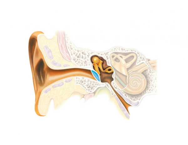
CSD 3103 anatomy of speech and hearing mechanisms Hearing mechanisms Fall 2008. The Inner Ear. The major divisions of the inner ear. The vestibular system The cochlea The osseous or bony labyrinth The membranous labyrinth. Location of the inner ear within the skull.

E N D
CSD 3103anatomy of speech and hearing mechanismsHearing mechanismsFall 2008 The Inner Ear
The major divisions of the inner ear • The vestibular system • The cochlea • The osseous or bony labyrinth • The membranous labyrinth
The cochlea Modiolus Internal auditory meatus Auditory nerve Facial nerve
The cochlea • Scala vestibuli • Scala tympani • Scala media • Helicotrema • Perilymph
The membranous labyrinth The membranous labyrinth in relation to the bony labyrinth
The membranous labyrinth Endolymph Semicircular canals Utricle Saccule Scala media
The membranous labyrinth Ampullae Crista ampularis Ductus Reuiens
Cross section of the cochlea Eighth Nerve Vestibular branch Acoustic branch Base Apex Modiolus Helicotrema Scala Vestibuli, Tympani and Media Basilar Membrane Reissner’s Membrane Spiral Lamina
Cochlear partitions An “unrolled” cochlea, showing the relationships among the three scalae
Path of vibration through the cochlea Vibrations are transmitted across scala media into scala tympani
Mass-action flow through the cochlea Inward movement of the stapes footplate causes perilymph to flow up scala vestibuli, through the helicotrema, and down scala tympani, where the round window is distended
Displacement of the basilar and tectorial membranes The vertical displacement of the basilar and tectorial membranes produces a shearing force on the cilia of the hair cells
Basilar membrane displacement Relative motion between the basilar and tectorial membranes places a shearing force on the stereocilia so they are bent away from the modiolus when the cochlear duct is displaced upward
Movement of the basilar membrane Schematic representation of the cochlea with the vestibule cut away. Arrows show the effects of a compressional sound wave produced by the stapes. Example of the position of a portion of the basilar membrane in 3 successive instants during sinusoidal stimulation.
Traveling wave Characteristics of the traveling wave generated by a 200 Hz stimulus peaking at a distance of about 29mm from the base of the cochlea
The traveling wave Schematic of amplitude patterns of traveling waves for sinusoids of various frequencies
Organization of the cochlea Tonotopic organization relates to the placement of auditory neurons in a particular structure (in this case, the cochlea) according to their responsiveness to specific frequencies