Download
1 / 44
480 likes | 702 Views
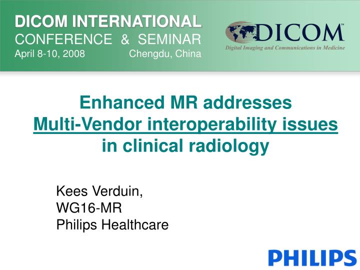
Enhanced MR addresses Multi-Vendor interoperability issues in clinical radiology. Kees Verduin, WG16-MR Philips Healthcare. Problem Statement. Multi Dimensional datasets create problems in a multi-vendor environment. Content. Enhanced MR Features /Benefits Multi–dimension storage

E N D
Enhanced MR addresses Multi-Vendor interoperability issues in clinical radiology Kees Verduin, WG16-MR Philips Healthcare
Problem Statement • Multi Dimensional datasets create problems in a multi-vendor environment.
Content • Enhanced MR Features /Benefits • Multi–dimension storage • From Chaos to Structure • Dimension Module • Clinical examples • Implementation scenarios • Conclusion
A new Standard, since 2003 Enhanced DICOM (e.g. for MR,CT,XA,PET,..) with DimensionModule
Enhanced MR Features Dimension Module Real World Values Multi-frame Spectroscopy Palette Color Raw Data Multi-stack
Enhanced MR Benefits • Transport Performance through new Multi-Frame concept • Better scan information new attributes for new scan techniques • Better Interoperability through more strict attribute rules • Context Information • Dimension Module • From “chaos” to “structure”
This is how these images were scanned ! 5 4 3 2 1 1 2 3 4 5
This is how these images were scanned ! 5 4 3 2 1 1 2 3 4 5
This is how these images were scanned ! 5 4 3 2 1 1 2 3 4 5
This is how these images were scanned ! 5 4 3 2 1 1 2 3 4 5
This is how these images were scanned ! 5 4 3 2 1 1 2 3 4 5
How to store the images ? • for easy retrieval • for consistent display • for optimal interpretation of parameters
Where to find the relevant data ? • DICOM images are sent as separate objects. • The relationship between the objects must be searched in Series attributes in these objects: • Like : Series Number, Series Description, Protocol Name, Sequence Name. • Despite 25 years of Image Format standardization, useful values for these attributes are not standardized ! • It will not be easy, if not impossible, to solve this in a standard. • Interpretation of image information is especially difficult when the objects are from different series. • Also when the relevant attributes themselves do not exist in the DICOM standard…. (private attributes like “number of .. “!)
From “Chaos” to “Structure” • Before “Enhanced MR” there was no standard way to solve this. • Now Enhanced DICOM for MR (and also for CT/XA/RF/PET etc..) can structure a dataset by: • packaging all images in one object and • using specific and better standardized attributesfor the organization. • The Dimension Module can disclose the organization of the dataset to the receiving application.
Where is the structure needed ? • In fact, this affects ALL Workstation SW implementations ! Note: in this presentation most examples are from MR, but similar problems exist for other modalities.
What should be done ? • There was a problem for (MR) users in a multi-vendor environment. • This was specifically a dimension problem, that has increased in importance over time. • Since 5 years there is already a solution ! • This is only slowly adopted by workstations and PACS vendors. • Users should update their workstations and PACS systems to support Enhanced DICOM.
MR creates, conceptually, a Multi-Dimensional Image dataset • Combinations of changing parameters create a whole space of images, which is defined by different dimensions. (Typical numbers) Echo (1-20) Time (30-10.000) Position (1-1000) Diffusion b-Value (3-10) Image type (1-3) Any parameter (x)
MR creates, conceptually, aMulti-Dimensional Image dataset • MR Scanners and MR Workstations from single vendors have dealt with this, in the classic DICOM MR Images. • The penalty was: • the use of private attributes for identificationand • vendors using different strategies for separating images into separate series.
Why was this not so much a problem in the past? • Most users had scanners and workstations from one vendor. • The storage choices were known by their own workstation SW Engineers. • The third-party workstation vendors had to reverse-engineer the separation strategies.
Increasing importance due to larger Enterprises • More and more hospitals are merging into bigger Healthcare Enterprises. • Users have more scanners and workstations from different vendors. • New Independent Workstations • New Independent PACS systems
Multiple images of the same slice,most likely in different series. • This leads to display problems for reading radiologists: • How can I see the values of the parameters that make the images different ? • How does my workstation organize the images that belong together ? • Why is it different for every scanner vendor ?
Enhanced DICOM, MultiFrame with Dimension Modules • All images are stored as “frames” in one image object. • Attributes may change from frame to frame. • Attributes that are important for the display order of the frames, will be structured in the Dimension Module by the creator. • This provides an explicit display scenario for the frames in the Enhanced (MR) object. • This scenario can be used by viewing systems, even without specific (MR) application knowledge. • It is vendor independent.
Clinical example: Perfusion One slice at many time points time
time Signal Clinical example: Perfusion One slice at many time points
Multi dimensional datasetsOrganization Attributes Example : Multi-slice –Multi-time acquisitions: • Every Frame has: • a slice order attribute value (“In-Stack Position Number”) and a time order attribute value (“Trigger Delay Time”). • These attributes are used as index in the Dimension Module. • The Dimension Module gives herewith the default display order
In-Stack Position Number Time One Enhanced MR Image object
Enhanced MR, supports the Multi-Dimension structure • All images stay together in one object. • The Dimension Module describes all relevant dimensions and indicates the priority for the display order. • A dedicated workstation may deviate from the suggested image ordering, by moving lower ranked dimensions upwards.
5 4 3 2 1 1 2 3 4 5 Total body Imaging requiresDimension Attributes Use‘Stack-ID’ and ‘In-Stack Position Number’ for correct display
Other Examples:Diffusion Imaging “Diffusion b-values” from 0 to 8000 and an ADC image
Other Examples: Cardiac Cine Loops Enables automatic multi-slice / multi-phase display, even for standard workstations
Implementation Roadmap • What if not all your systems are ready for Enhanced DICOM ?
The Negotiation scenario PACS can not send and must cancel ! MR decides to send Enhanced object Archive MR Negotiate Negotiate Negotiate Workstation MR decides to send the Classic MR object Conclusion: One can not configure a PACS for Enhanced MR, as long as it is not supported in the diagnostic workflow.
Back to Square one ? MR decides to send the Classic MR object PACS decides to send the Classic MR object Archive MR Negotiate Negotiate Workstation Conclusion: No support for Enhanced MR (and “chaos” continues)
Negotiate MR decides to send the Classic MR object Action required:Adapt the workstations ! PACS decides to send Enhanced object! PACS can not send and must cancel ! MR decides to send Enhanced object Archive MR Negotiate Negotiate Workstation Action: Replace the blocking (part of the) Workstations
Conclusions • All Workstations and all Image Managers should support and use Enhanced DICOM SOP Classes with the Dimension Module. • Enhanced (MR) objects exist worldwide.>1600 MR systems are waiting to send their data. • Only then, the clinical users can benefit from the other features of Enhanced Objects
Summary • Enhanced DICOM solves Multi Vendor Dimension problems. • Workstation and PACS vendors must implement this. • Hospitals must invest in upgrades to fully benefit.
Further Information ? See: Henri Matthijssen with his poster about the Philips Implementation of Enhanced MR