Download
1 / 85
860 likes | 884 Views
Explore the crucial skeletal features of the pelvis, hip, and thigh, including bones, joints, ligaments, and musculature. Learn about the structure and function of the pelvis, hip joint, femur, and associated bursae. Gain insights into the kinematics, muscles, nerves, blood vessels, and prevention strategies for hip injuries.

E N D
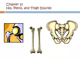
Pelvis, Hip, and Thigh Conditions Chapter 17
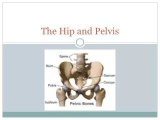
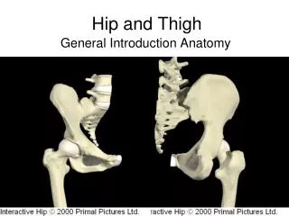
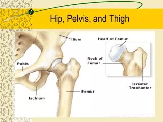
Pelvis • Function • Protects organs • Transmits loads between trunk and lower extremity • Provides site for muscle attachments • 4 fused bones • Sacrum • Coccyx • Innominate bones • Ilium, ischium, and pubis
Pelvis (cont.) • SI joint • Critical link between the two pelvic bones • Strong ligamentous support • Sacrococcygeal joint • Fused line symphysis united by a fibrocartilaginous disc • Pubic symphysis • Interpubic disc located between the two joint surfaces
Bony Structure of Thigh • Femur • Weakest at femoral neck • Angle of inclination • Angle of depression formed by a line drawn through the shaft of femur and a line passing through the long axis of femoral neck • Approximately 125 in the frontal plane • 125 coxa valga • 125 coxa vara
Bony Structure of Thigh (cont.) • Femur • Angle of torsion • Relationship between femoral head and femoral shaft in transverse plane • Approximately 12 • 12 anteversion • 12 retroversion
Hip Joint • Head of femur and acetabulum of pelvis • Ball and socket joint • Very stable
Hip Joint Capsule • Completely surrounds joint, attaching to the labrum of the acetabular socket • Passes over a fat pad internally to join to the distal aspect of femoral neck • Zona orbicularis
Ligaments of Hip Joint • Iliofemoral ligament • Limits hyperextension • Pubofemoral ligament • Limits abduction and hyperextension • Ischiofemoral ligament • Limits extension
Femoral Triangle • Borders • Inguinal ligament—superior • Sartorius—lateral • Adductor longus—medial • Contents • Femoral nerves • Femoral artery • Femoral vein
Bursae • Iliopsoas • Reduces friction between iliopsoas and articular capsule • Deep trochanteric bursa • Provides cushion between greater trochanter and gluteus maximus at its attachment to iliotibial tract • Gluteofemoral bursa • Separates gluteus maximus from origin of vastus lateralis • Ischial bursa • Weight-bearing structure during sitting • Cushions ischial tuberosity where it passes over gluteus maximus
Q-Angle • Angle between line of resultant force produced by quadriceps and line of patellar tendon • Males 13°; females 18°
Nerves • Lumbar plexus • Femoral nerve • Obturator nerve • Sacral plexus • Sciatic nerve
Blood Vessels • External iliac • Femoral • Deep femoral • Femoral circumflex • F16.10
Kinematics (cont.) • Hip flexors • Iliopsoas, pectineus, rectus femoris, sartorius, and tensor fascia latae • Two-joint muscles • Rectus femoris—active during hip flexion and knee extension • Sartorius—active during hip flexion and knee extension • Hip extensors • Gluteus maximus and hamstrings (biceps femoris, semitendinosus, and semimembranosus) • Hamstrings—two-joint; hip extension and knee flexion
Kinematics (cont.) • Hip abductors • Gluteus medius, gluteus minimus • Active in stabilizing pelvis during single-leg support and during support phase of walking and running • Hip adductors • Adductor longus, adductor brevis, and adductor magnus
Kinematics (cont.) • Lateral rotators • Piriformis, gemellus superior, gemellus inferior, obturator internus, obturator externus, and quadratus femoris • Lateral rotation of femur of swinging leg accommodates lateral rotation of pelvis during stride • Medial rotators • Gluteus minimus • Tensor fascia latae, semitendinosus, semimembranosus, gluteus medius, and adductors
Kinetics • Body weight places compression on hip, as does tension in hip muscles • Forces are less during standing than with running and walking • Forces translated through the lower extremity; result ↑ compression on hip
Prevention • Protective equipment • Hip joint well protected but iliac and pelvis need protection • Thigh • Physical conditioning • Shoes • Cushion forces
Contusions • Hip pointer • Mechanism:direct blow to iliac crest • Common—anterior or lateral portion of crest • Often from improperly fitting (or absent) hip pads
Contusions (cont.) • S&S • Point tenderness; swelling; ecchymosis • Individual prefers slightly forward flexed position to relieve tension of abdominals and iliopsoas • Antalgic gait with shortened swing phase • ↑ pain with active trunk flexion and active hip flexion • Pain with coughing, laughing, breathing • Abdominal muscle spasm • Management: standard acute; rest; protect with hard-shell pad for return to activity
Contusions (cont.) • Quadriceps contusion • Mechanism: direct blow • Common – anterolateral thigh • S&S • Transitory loss of function • With continued play, progressively stiffer and unresponsive • ↑ pain with active knee extension and hip flexion • Limited AROM due to pain; knee flexion limited actively and passively
Contusions (cont.) • Management: • Standard acute; with knee in maximum flexion • Hard-shell pad for return to activity • Physician referral if myositis ossificans orcompartment syndrome is suspected
Contusions (cont.) • Myositis ossificans • Develops secondary to single significant blow or repetitive blows to same area • Evident on radiograph 3–4 weeks after injury
Contusions (cont.) • S&S • Warm, firm, swollen thigh; 2–4 cm larger • Palpable, painful mass may limit passive knee flexion to 20–30° • Active quadriceps contractions and straight leg raises—difficult • Management: standard acute; physician referral • Self-limiting injury • Maturation—6–12 months
Contusions (cont.) • Compartment syndrome • Neurovascular compression • Due to uncontrolled internal bleeding and swelling • S&S • Progressive, severe pain with passive motion and isometric contraction of quadriceps • pressure →↓ femoral sensation and motor weakness; distal pulse and capillary refill may be normal • Management: ice (no compression); immediate physician referral
Bursitis • Mechanism • Excessive friction or shear forces due to overuse • Posttraumatic bursitis from direct blows that cause bleeding in the bursa • Greater trochanteric bursitis • Influence of Q-angle
Bursitis (cont.) • S&S • Burning or aching over or posterior to greater trochanter • Aggravated with: • Hip abduction against resistance • Hip flexion and extension on weight bearing • Referred pain—lateral aspect of the thigh
Bursitis (cont.) • Iliopsoas bursitis • Pain medial and anterior to joint; cannot be easily palpated • pain with passive hip rotation; resisted hip flexion, abduction, and external rotation • Ischial bursitis • Pain aggravated by prolonged sitting and uphill running, • Point tenderness directly over ischial tuberosity • pain with passive and resisted hip extension
Bursitis (cont.) • Bursitis management • Standard acute; deep friction massage; NSAIDs; stretching program for involved muscle • On-going prevention: biomechanical analysis; technique analysis
Bursitis (cont.) • Snapping hip syndrome • Causes: intra- and extra-articular (refer to Box 15.2) • Types • External—IT band or gluteus maximus snapping over greater trochanter during hip flexion → trochanteric bursitis • Internal—iliopsoas snaps over structures deep to musculotendinous unit (e.g., iliopsoas bursa) • Intra-articular—lesions of the joint (e.g., labral tear)
Bursitis (cont.) • S&S • Snapping sensation heard or felt during hip motion, especially with lateral rotation and flexion while balancing on one leg • Iliopsoas bursa affected—snapping in medial groin • Management: NSAIDs; rehabilitation program to address specific deficits
Hip Sprains and Dislocations • Mechanism • Violent twisting actions • With hip and knee flexed to 90°, force through shaft of femur
Hip Sprains and Dislocations (cont.) • S&S • Mild/moderate: pain with internal rotation • Severe: intense pain; inability to move hip • Position of flexion and internal rotation • Management • Mild/moderate—standard acute • Severe—activate EMS; immobilize in position found; assess distal vascular integrity; monitor and treat for shock; NPO
Strains • Mechanism • Explosive movements • Tensile stress from overstretching • Muscles • Quadriceps • Typically rectus femoris
Strains (cont.) • Hamstrings • Initial swing—flex knee; late swing—eccentrically contract to decelerate knee extension and re-extend hip in prep for stance phase • Overemphasis on stretching without strengthening • Strength imbalance • Adductors • Common with quick change of direction and explosive propulsion and acceleration • Strength imbalance
Strains (cont.) • S&S • Point tender with palpable spasm • Possible palpable defect/divot • Ecchymosis may or may not be present • Pain with AROM; pain with PROM (muscles placed on stretch)
Strains (cont.) • Piriformis strain • In some individuals, sciatic nerve passes through or above piriformis, subjecting nerve to compression from trauma, hemorrhage, or spasm
Strains (cont.) • S&S • History of prolonged sitting, overuse, recent ↑ in activity, or buttock trauma • Dull ache in midbuttock—worse at night • Numbness or weakness may extend down posterior leg • ↑ pain or weakness during: • Passive hip flexion, adduction, and internal rotation • Active hip external rotation • Resisted hip external rotation
Strains (cont.) • Predisposing factors • Beginning of season – too much too soon • Fatigue • History of strains; reinjury common • Restricted flexibility of involved muscle group • Management: standard acute; restrict weight bearing if unable to assume normal gait
Vascular and Neural Disorders • Legg-Calvé-Perthes disease • Avascular necrosis of proximal femoral epiphysis • Seen esp in males ages 3–8 • Osteochondrosis - femoral head • S&S • Gradual onset of limp and mild hip or knee pain of several months in duration • Pain -activity related • ROM in hip abduction, extension, and external rotation due to spasm in hip flexors and adductors
Vascular and Neural Disorders (cont.) • Venous disorders • Direct blow may damage a vein causing • Thrombophlebitis • Superficial thrombophlebitis (ST) • Deep venous thrombosis (DVT) • Phlebothrombosis
Vascular and Neural Disorders (cont.) • S&S • ST—acute, red, hot, palpable, tender cord in course of a superficial vein • Extension of ST to deep veins—via proximal long and short saphenous veins to common femoral and popliteal veins, respectively • Management: anticoagulant therapy; external support (e.g., compression stockings); therapeutic exercise