Download
1 / 32
430 likes | 1.67k Views
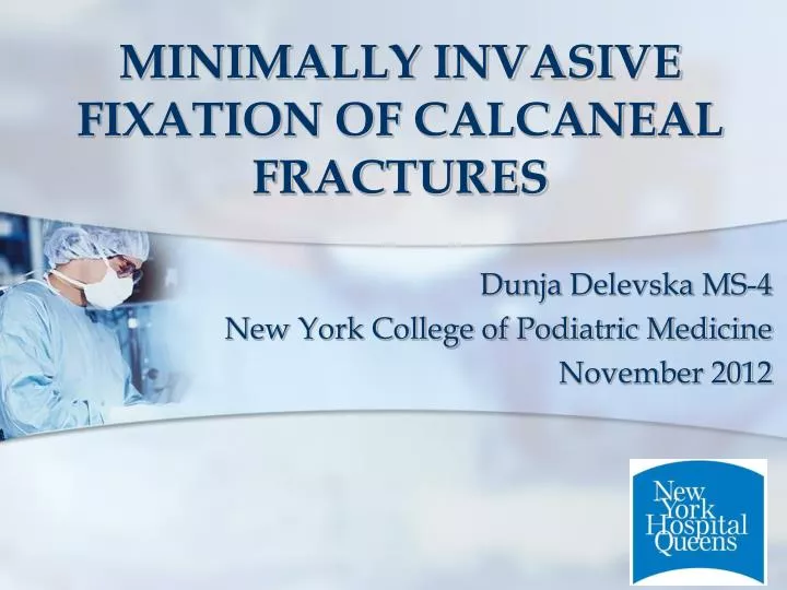
MINIMALLY INVASIVE FIXATION OF CALCANEAL FRACTURES. Dunja Delevska MS-4 New York College of Podiatric Medicine November 2012. OUTLINE. Imaging Classification Management Literature . PRESENTATION.

E N D
MINIMALLY INVASIVE FIXATION OF CALCANEAL FRACTURES DunjaDelevska MS-4 New York College of Podiatric Medicine November 2012
OUTLINE • Imaging • Classification • Management • Literature
PRESENTATION 35 y.o. male patient presents to ED with cc of foot pain b/l. S/p fell in a pothole while texting No open fracture Patient is AAO x3 Denies LOC
IMAGING DP X-Ray of the foot Lateral of the foot and ankle Bohler’stuberosity joint angle Crucial angle of Gissane • Calcaneocuboid joint involvement • Medial subluxation of talus at TNJ – severe fractures
IMAGING (cont.) Right foot X-Rays Evaluation: Bohler’s angle – normal Gissane angle – normal Increased sclerosis in calcaneal body on MO No displacement
IMAGING (cont.) Right foot CT Displaced?
IMAGING (cont.) Left foot CT Displacement? Comminution?
IMAGING (cont.) Spine X-Ray L2 compression fracture
CLASSIFICATION ROWE CLASSIFICATION Type Ia – fracture of the inferior calcanealtuberosity
CLASSIFICATION (cont.) ROWE CLASSIFICATION Type Ib – sustentaculumtali fracture
CLASSIFICATION (cont.) SANDERS CLASSIFICATION IIIAC Medial edge of posterior facet (C) Through posterior facet (A)
CLASSIFICATION (cont.) • Medial primary fracture line • Increased Sanders class • DecrasedBohler’s angle • CCJ involvement • Anterior and middle facet involvement • increased severity Silhanek et al. The Effect of Primary Fracture Line Location on the Pattern and Severity of IntraarticularCalcaneal Fractures: A Retrospective Radiographic Study. J Foot Ankle Surg. 2006 Jul/Aug; 45(4): 211-219
CLASSIFICATION (cont.) Centrally depressed fragment Increased Gissane’s angle
MANAGEMENT What should be addressed? • Immobilization of non-displaced fracture • Reduction of joint depression and correction of Gissane’s angle and cortical strut • Restoration of height and width of calcaneus • Anatomic alignment of articular surfaces • Heel varus
MANAGEMENT (CONT.) Right foot non-displaced fracture • Closed reduction (if necessary) • Immobilized with below-the-knee cast • Cast in mild inversion to take stress off the fracture • Early ROM of hallux to prevent adhesion of FHL to the sustentaculum during healing
MANAGEMENT (CONT.) Left foot comminuted fracture • General anesthesia with popliteal block • Patient placed in lateral decubitus position with left side up • Minimal incision reduction with internal fixation • Fracture fragments anatomically reduced • Restore posterior facet and angle of Gissane • Four 4.0 cannulated screws
MANAGEMENT (CONT.) POPLITEAL BLOCK
MANAGEMENT (CONT.) MODIFIED OLLIER INCISION
MANAGEMENT (CONT.) ORIF – Four 4.0 cannulated screws
MANAGEMENT (CONT.) What was accomplished?
MANAGEMENT (CONT.) Correction of Bohler’s angle
MANAGEMENT (CONT.) Correction of angle of Gissane
LITERATURE REVIEW The skin pliability and wrinkles are evaluated until the skin appears as it does here. Bergin et al. Inpatient soft tissue protocol and wound complications in calcaneus fractures. Foot Ankle Int. 2012 Jun;33(6):492-7.
LITERATURE REVIEW (CONT.) Mostafa et al. Surgical treatment of displaced intra-articularcalcaneal fracture using a single small lateral approach. Strategies Trauma Limb Reconstr. 2010 Aug;5(2):87-95. Epub 2010 Mar 9.
LITERATURE REVIEW (CONT.) Stulik et al. Minimally-invasive treatment of intra-articular fractures of the calcaneum. J Bone Joint Surg Br. 2006 Dec;88(12):1634-41.
LITERATURE REVIEW (CONT.) Stulik et al. Minimally-invasive treatment of intra-articular fractures of the calcaneum. J Bone Joint Surg Br. 2006 Dec;88(12):1634-41.
LITERATURE REVIEW (CONT.) Minimally invasive technique of Forgon and Zadravecz Tomesen et al. Treatment of displaced intra-articularcalcaneal fractures with closed reduction and percutaneous screw fixation. J Bone Joint Surg Am. 2011 May 18;93(10):920-8.
LITERATURE REVIEW (CONT.) DeWall et al. Percutaneous reduction and fixation of displaced intra-articularcalcaneus fractures. J Orthop Trauma. 2010 Aug;24(8):466-72.
SUMMARY • Inspect for concomitant injuries and ensure patient is medically stable before definitive treatment / Inspect soft tissue • Obtain proper imaging that would aid in treatment decision making • Classify fractures to aid in surgical decision making and prognosis • Ensure proper anatomic alignment and employ stable/biologic internal fixation
REFERENCES • Silhanek et al. The Effect of Primary Fracture Line Location on the Pattern and Severity of IntraarticularCalcaneal Fractures: A Retrospective Radiographic Study. J Foot Ankle Surg.2006 Jul/Aug; 45(4): 211-219 • Bergin et al. Inpatient soft tissue protocol and wound complications in calcaneus fractures. Foot Ankle Int. 2012 Jun;33(6):492-7. • Mostafa et al. Surgical treatment of displaced intra-articularcalcaneal fracture using a single small lateral approach. Strategies Trauma Limb Reconstr. 2010 Aug;5(2):87-95. Epub 2010 Mar 9. • Stulik et al. Minimally-invasive treatment of intra-articular fractures of the calcaneum. J Bone Joint Surg Br. 2006 Dec;88(12):1634-41. • Tomesen et al. Treatment of displaced intra-articularcalcaneal fractures with closed reduction and percutaneous screw fixation. J Bone Joint Surg Am. 2011 May 18;93(10):920-8. • DeWall et al. Percutaneous reduction and fixation of displaced intra-articularcalcaneus fractures. J Orthop Trauma. 2010 Aug;24(8):466-72.