Download
1 / 6
70 likes | 535 Views
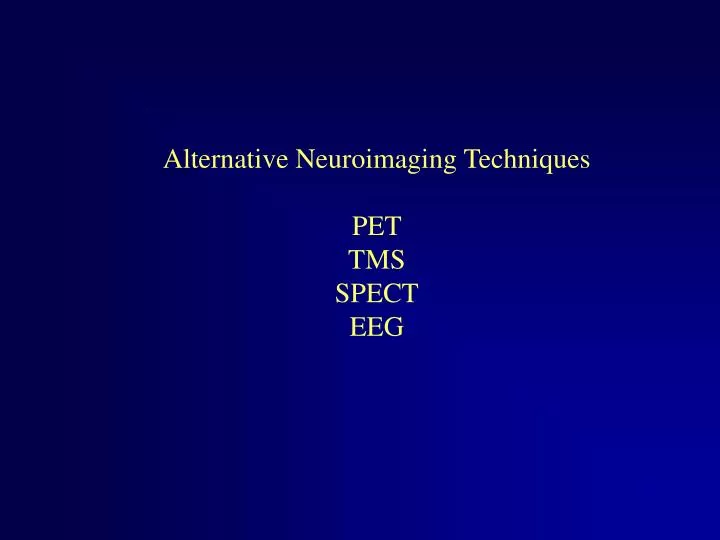
Alternative Neuroimaging Techniques PET TMS SPECT EEG. Positron Emission Tomography (PET). Relies on injection of a radioactive isotope to measure cerebral blood flow.

E N D
Positron Emission Tomography (PET) Relies on injection of a radioactive isotope to measure cerebral blood flow. Whereas fMRI images rely on the interaction of multiple factors (different tissue proton densities, relaxation times, a combination of CBF, CBV, CRMO and CRMGl) PET relies only on only one factor for a given experiment (e.g., usually CBF – but can be CBV or CRMO)
Transcranial magnetic stimulation (TMS) Typically an interruption of function – creating temporary lesions in the healthy brain. Great for pinpointing regions involved in specific components of tasks or for mimicking neurological disorders. Single vs. rapid-pulse TMS – inherent dangers in rapid-pulse TMS Poor spatial resolution – vitamin E tablets and MRI help! (and magnetic dipole modeling as in VEPs)
Single Photon Emission Computed Tomography (SPECT) SPEC T PET Less spatial resolution than PET – but far less expensive Often used for early detection of dementias – evidenced by hypoperfusion in a given area http://imasun.lbl.gov/~budinger/medTechdocs/SPECT.html
Electro-encephalogram (EEG) Scalp potentials – EEG used clinically but from this we can get visually evoked potentials (VEPs) and the like. Dipole modeling used to locate the source of the VEP Requires many trials – poor spatial but great temporal resolution. 128 electrode array
Combinations fMRI and single cell in monkeys (Logothetis) Non-pherous EEG recordings with fMRI in humans TMS and PET (Paus, 1999; Desmurget et al. 1999)