Download
1 / 1
10 likes | 85 Views
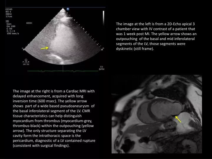
The image at the left is from a 2D-Echo apical 3 chamber view with IV contrast of a patient that was 1 week post MI. The yellow arrow shows an outpouching of the basal and mid inferolateral segments of the LV, those segments were dyskinetic (still frame).

E N D
The image at the left is from a 2D-Echo apical 3 chamber view with IV contrast of a patient that was 1 week post MI. The yellow arrow shows an outpouching of the basal and mid inferolateral segments of the LV, those segments were dyskinetic (still frame). The image at the right is from a Cardiac MRI with delayed enhancement, acquired with long inversion time (600 msec). The yellow arrow shows part of a wide based pseudoaneurysm of the basal inferolateral segment of the LV. CMR tissue characteristics can help distinguish myocardium from thrombus (myocardium-grey, thrombus-black) within the outpouching(yellow arrow). The only structure separating the LV cavity form the intrathoracic space is the pericardium, diagnostic of a LV contained rupture (consistent with surgical findings).