Download
1 / 44
440 likes | 613 Views
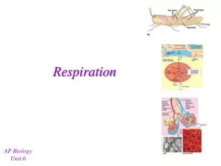
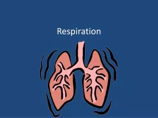
Respiration. Learning objectives. To outline the respiratory system To define lung volumes and capacities. To describe the mechanisms of respiration. To describe the mechanisms of oxygen and carbon dioxide transport. To describe the control of respiration. The air passage. Nasal cavity.

E N D
Learning objectives • To outline the respiratory system • To define lung volumes and capacities. • To describe the mechanisms of respiration. • To describe the mechanisms of oxygen and carbon dioxide transport. • To describe the control of respiration.
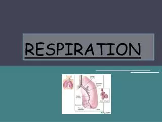
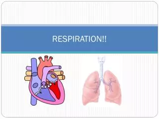
The air passage Nasal cavity Pharynx Larynx Trachea Bronchi Lungs
Nasal cavity • Air enter the nasal cavity through the two nostrils; • On route, the air is ~ warmed ~ moistened ~ filtered by the hairs lining the cavity
Pharynx • It leads to two passages: ~ trachea ~ oesophagus • Epiglottis closes the trachea to prevent food from entering the air-way during swallowing.
Larynx • It is at the upper end of the trachea. • It is composed of 5 pieces of cartilage: the vocal cords. • The vocal cords vibrate to produce sound whenever air tension passes over them.
Trachea • A flexible pipe-like structure supporting by C-shaped ring of cartilage. • Mucosa secreting mucus is lining in the inner wall of trachea. • Cilia are found on the mucosa. • Movement of cilia remove any foreign object or mucus from the trachea.
Bronchi • The trachea divides to form two bronchi: ~ right bronchus right lungs ~ left bronchus left lungs • The bronchus divides repeatedly into bronchioles inside each lungs. • The bronchioles lead into air sacs eventually.
Lungs • It is the place for gaseous exchange: exchange of carbon dioxide and oxygen between environment and organism. • Within each lung: Bronchus smaller bronchioles air sacs & alveolus
Features of alveoli • Large surface area • Thin wall • Moist surface • Rich supplied with blood capillaries
Pleural membrane • It is a sheet of smooth membrane. • It covers the outside surface of the lungs and the inside surface of the thorax. • It consists of two layers: ~ inner pleura ~ outer pleura • Pleuralcavity is present between the pleurae. • Pleural fluid fills the pleural cavity. • Pleural fluid reduce friction between pleurae during breathing.
Ventilation • It is the passage of air into and out of the respiratory tract. • Breathing is the act. • It consists of ~ inspiration( breathe in) ~ expiration(breathe out)
Accessories for ventilation • Diaphragm ~ fibrous sheet of tissue ~ dome shape when it relaxes (expiration) ~ flatten when it contracts(inspiration) • Intercostal muscle ~ muscle between ribs • Anterior abdominal wall
Inspiration • Diaphragm: contracts and flattens • Anterior abdominal wall: relaxes • Thoracic volume: • Pressure in pleural cavity: • Movement of air: into the lungs • Shape of the lungs : inflated
Expiration • Diaphragm: relaxes and becomes dome shape • Anterior abdominal wall: contracts • Thoracic volume: • Pressure in pleural cavity: • Movement of air: forced out of the lungs • Shape of lungs: deflated
Lung volume and capacitites • Tidal volume • Inspiratory reserve volume • Expiratory reserve volume • Vital capacity • Residual volume • Dead space • Total lung capacity
Tidal volume • The amount of air movesin or out of the lungs with a singlebreath at rest. • It is about 0.5 L.
Inspiratory reserve volume • The amount of air that can be taken into the lungs over and above the tidal volume. • It is about 1.5 L.
Expiratory reserve volume • The extra amount of air that can be forced out after a normal expiration. • It is about 1.5 L.
Vital capacity • The greatest volume of air that can be expired after a maximum inspiratory effort. • It is about 3.5L.
Residual volume • The amount of airremain in the lung even after forced expiration. • It is about 1.5L.
Dead space • The volume of inspired air which cannot reach lungs for gaseous exchange and remains in the respiratory tract. • It is about 0.15L.
Total lung capacity • Vital capacity + residual volume. • It is about 5 L.
Transport of gases • Haemoglobin is the most common example of respiratory pigment. • Haemoglobin is an iron-containing pigment (haem group). • Haemoglobin is confined to red blood cells
Transport of oxygen Low O2 tension Hb + O2 HbO2 + O2 HbO4 + O2 HbO6 + O2 High O2 tension
Transport of oxygen • At the lungs: ~ amount of oxygen (PO2) is huge ~ oxygen binds to haemoglobin(Hb) to form oxyhaemoglobin ~ nearly 95% of Hb is saturated with oxygen ~ 100% saturation of haemoglobin is rarely achieved
Transport of oxygen • At tissue: ~ amount of oxygen(PO2) is limited ~ haemoglobin cannot binds with the oxygen ~ oxyhaemoglobin dissociates and release oxygen to the tissue cells
S-shaped curve • The oxygen dissociation curve is S-shaped (sigmoid). • The shape of haem group distorts as O2 combines with the Hb. • The distortion enhances further attachment of O2 to Hb. • The more the O2 that have been loaded, the faster further attachment.
S-shaped curve • The first O2 is released to the tissues very rapidly. • But the second, third and fourth O2 are given up much less readily. The S-shaped oxygen dissociation curve provides the mechanism for fast loading and unloading of oxygen.
Factors affecting the oxygen dissociation curve I • Acidity (Carbon dioxide concentration) ~ PCO2, efficiency at taking up O2 by Hb, but efficiency at releasing it. e.g. At muscle and liver ~ high respiration rate of the cells ~ rapid release of O2 from the blood supplying them
Factors affecting the oxygen dissociation curve II • Temperature ~ blood temperature, affinity of Hb for O2 unloading of O2 from the Hb e.g. oxygen dissociate from Hb efficiently and rapidly in endotherms(warm blooded animal) than in ectotherms(cold blooded animal).
Factors affecting the oxygen dissociation curve III • Altitude ~ altitude, volume of O2 in the atmosphere, affinity of Hb for O2 • Size of animal ~ size, affinity of affinity for O2 unloads it more readily
Factors affecting the oxygen dissociation curve IV • Fetal haemoglobin ~ the fetal haemoglobin has a higher affinity for O2 than maternal haemoglobin; ~ the fetal blood pick up O2 from maternal blood across the placenta readily.
Factors affecting the oxygen dissociation curve V • Carbon monoxide ~ the affinity of Hb for carbon monoside is several hundred times as great as it is for O2; ~ Hb will combines with any carbon monoxide available in preference to O2 As little as 0.1% carbon monoxide is dangerous, it causes asphyxiation.
Factors affecting the oxygen dissociation curve VI • Myoglobin ~ It release O2 only when the O2 concentration falls very low; ~ myoglobin stores O2 in the muscle and releases in an emergency case.
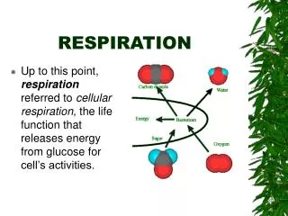
Transport of carbon dioxide • In plasma (5%) ~ carbonic acid forms in this process ~ the acid influences the plasma pH greatly • Enter red blood cells(95%)
Control of ventilation • Breathing centre • Chemoreceptors • Baroreceptors • Stretch receptors • Voluntary control • Effect of carbon dioxide on breathing rate • Effect of oxygen
Breathing centre • It is in the medulla oblongata of the brain. • It sends impulses to the intercostal muscle and diaphragm. • The muscle respond by bringing thorcic movements. • It consists of inspiratory centre and expiratory centre.
Chemoreceptors • They are located in the carotid and aortic bodies of the blood system. • When concentration of carbon dioxide , they discharge impulses to the respiratory centre.
Baroreceptors • They are located at the carotid artery and aorta. • They detect the change of blood pressure. • Blood pressure, the baroreceptor discharge impulse to respiratory centre, rate and depth of breathing.
Stretch receptors • They are located in the wall of alveoli. • Lung expand stimulates the stretch receptors impulses to the respiratory centre switches off the inspiratory centre.
Voluntary control • The rate and depth of breathing can be controlled by our mind. • Cerebrum sends impulses to the breathing centre.
Effect of carbon dioxide acidity ( carbon dioxide concentration) Respiratory centre send impulses Diaphragm & intercostal muscle in general, PCO2, fast & large ventilation
Effect of oxygen • The respiratory centre is insensitive to oxygen concentration in blood. • The chemoreceptors only respond to serious decrease in oxygen concentration. • The regulation of breathing depends primarily on accumulation of carbon dioxide and not only lack of oxygen.