Download
1 / 41
730 likes | 2.06k Views
Basics of Chest X-Ray. AFAMS Residency Orientation April 16, 2012. Outline. CXR Basics Types of CXR PA vs. AP Films Obtaining Images Systematic method to reading CXR Common Signs Examples. Chest X-ray (CXR) Basics. A standard chest X-ray consists of a PA Image Lateral Image

E N D
Basics of Chest X-Ray AFAMS Residency Orientation April 16, 2012
Outline • CXR Basics • Types of CXR • PA vs. AP Films • Obtaining Images • Systematic method to reading CXR • Common Signs • Examples
Chest X-ray (CXR) Basics • A standard chest X-ray consists of a • PA Image • Lateral Image • Images read together • AP for supine patients • Lots of information available on a CXR • Be systematic with your reading • Always compare to prior studies if possible
Basics of X-Rays • X-Rays are part of the light spectrum • Unlike visible light, x-rays pass through the human body • Pass through lungs without much interference • Difficult to pass through bones • Place film cassette on other side of patient and capture the shadow
Basics of X-Ray • Organs absorb X-rays differently and thus their shadow on the film is different • Bone: high absorption (film appears white) • Tissue: moderate absorption (film appears grey) • Air/Lungs: little absorption (film appears black)
Types of CXRs • PA and Lateral • Patient facing cassette • X-ray 6 feet away • Supine AP • X-ray 40 inches away • Magnifies anterior structures and pulmonary vasculature 101 cm 1.83 m
Comparing Chest X-rays Protocols PA AP Note heart enlarged, lung fields not as clear • Preferred method
PA Image • PA Film • Read as if patient is facing you (Patient’s left side is on the right of the X-ray)
Lateral Image • Obtained with patient’s left side against the cassette. • Minimizes heart silhouette magnification
Assessing Film Technique • Inspiration • Penetration • Rotation
Inspiration • Image should be at full inspiration • Diaphragm at level of 8-10 rib • Allows reader to see intrapulmonary structures Poor Inspiration mimics RML Infiltrate Same patient with proper inspiration
Penetration • Amount of radiation required for a quality image • PA film: should barely see thoracic spine disc spaces • Lateral: spine should appear darker as move cadually Examples of adequately penetrated images
Penetration Overpenetrated Underpenetrated
Rotation • Patient should be flat against the cassette • Rotation of the patient will alter appearance of mediastinum • Observe rotation by comparing location of clavicular heads • Should be equal distance from spinous process of thoracic vertebral bodies
Rotation Normal Rotated to the Right
Mass vs. Infiltrate Mass Infiltrate
Lobes and Fissures: PA Film A: Minor Fissure between RML and RLL B: Upper and lower boundaries of major fissures
Lobes and Fissures: Lateral A: Minor Fissure R Lung B: Major Fissure R Lung B: Major Fissure L Lung
How to Read an X-Ray Part 1 • Patient Data (Name, history, age, sex) • Technique (PA vs. AP, rotation, penetration, etc) • Trachea: midline or deviated, any masses? • Lungs: masses, infiltrates? • Costophrenic angles should be sharp (if not = effusions) • Silhouette signs, air-bronchograms, pulmonary edema • Pulmonary vessels: enlarged?
How to Read an X-Ray Part 2 • Hilar Region: masses or lymphadenopathy • Heart: enlarged, abnormal shape • Pleura: effusion, thickening, calcification • Bones: fractures or masses • ICU Films: looks for line and tube placement
How to Read an X-Ray Part 3 • It is best to focus on a small area of the film and then scan rather than look at the whole film at once
Signs: Silhouette Sign • Loss of lung/soft tissue interface caused by mass, fluid, or infiltrate in the normally air filled lung • Commonly applied to heart, aorta, chest wall, and diaphram borders with lung • Location of silhouette sign helps to localize pathology Lose Right Heart and Lung border = RML
Signs: Air Bronchogram • Tubular outline of an airway made visible by filling of the surrounding alveoli by fluid or inflammatory exudates • Causes • Pulmonary edema • Lung Consolidation • Severe Interstitial Disease • Neoplasm
Signs: Solitary Pulmonary Nodule • Can be innocuous or potentially fatal lung cancer • Always compare to prior films for growth • Nodules with irregular borders are suspicious
Conclusions • Lots of information in a chest x-ray • Always read the film in the same order • Never skip to the most prominent abnormality, you will miss a small (but potentially important finding) • Compare to priors if possible • We will finish with some examples of common pathology
Examples: Atelectasis • Collapse or incomplete expansion of alveoli • Causes: • Endobronchial lesions (mucous plug or tumor) • Extrinsic compression (mass, lymph node) • Peripheral compression (pleural effusion) • Linear density on CXR
Examples: Pulmonary Edema • Cephalization of pulmonary vessels (arrow) • Kerley B Lines • Peribronchial cuffing • “Bat Wing” Appearance • Increased Cardiac Size (arrow)
Examples: Pneumonia • Airspace disease and consolidation • CXR Findings • Airspace opacity • Lobar consolidation • Interstitial opacities
Differentiating Atelectasis from Pneumonia Atelectasis Pneumonia Normal or increased volume No shift Consolidation, air space process Not centered at hilum Air bronchograms • Volume Loss • Associated ipsilateral shift • Linear, wedge shaped • Apex at hilum • Air bronchograms
Examples: TB • TB can be seen as consolidation, cavitation, fibrosis, adenopathy, or pleural effusion depending on stage of infection
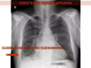
Examples: Pleural Effusions Fluid in Costophrenic Angle Blunting of Costophrenic Angles
Examples: Pneumothorax (PTX) • Air inside the thoracic cavity but outside the lung • PTX appears as air without lung markings in least dependent area of chest
Examples: Hemopneumothorax Lung Air Fluid
Examples: Interstitial Lung Disease • Hazy ground glass opacification • Volume Loss • Linear opacities bilaterally • “Honeycomb lung”
Examples: COPD and Emphysema • Diffuse hyperinflation • Flattened diaphragms • Increased retrosternal space • Bullae
Examples: Rib Fractures • Can you find the rib fracture?
Examples: Hiatal Hernia Gastric Bubble
Hilar Enlargement Enlarged Pulmonary Artery Hilar Adenopathy