Download
1 / 41
480 likes | 584 Views
RESPIRATION. Dr. Zainab H.H Dept. of Physiology Lec.6,7. Objectives. Describe the importance of ventilation/perfusion matching at the alveolar level in maintaining proper levels of systemic arterial blood gases.

E N D
RESPIRATION Dr. Zainab H.H Dept. of Physiology Lec.6,7
Objectives • Describe the importance of ventilation/perfusion matching at the alveolar level in maintaining proper levels of systemic arterial blood gases. • Explain how ventilation/perfusion matching is physiologically achieved in gravity fields operating on fluids (air/blood) of such different mass. • Generate an alveolar PO2-PCO2 diagram that identifies the three alveolar types, showing the continuum of ventilation/perfusion ratios. • Identify in vivo pulmonary reflexes that help to correct for vascular shunts or airway obstructions causing ventilation/perfusion disturbances.
Objectives (continue) • Determine the effect of exercise on V/Q ratio • Discriminate the Gas Exchange at Alveolar and Tissue Level • List the factors that affect the rate of gases diffusion in the blood
Pulmonary Circulation • In a fetus: • Pulmonary circulation has a higher vascular resistance, because the lungs are partially collapsed. • After birth, vascular resistance decreases: • Opening the vessels as a result of subatmospheric intrapulmonary pressure. • Physical stretching of the lungs. • Dilation of pulmonary arterioles in response to increased alveolar PO2.
Pulmonary Circulation (continued) • In adult the Rt. ventricle (like the left) has a COP of about 5.5 L per minute. • Thus blood flow through the pulmonary circulation =flow rate through the systemic circulation. • Driving pressure = 10 mmHg in pulmonary circulation = 1/10th of systemic circulation. • pulmonary vascular resistance is 1/10th the systemic vascular resistance • Thus, pulmonary circulation is a low-resistance, low-pressure pathway. • Less net filtration pressure than produced in the systemic capillaries avoids pulmonary edema.
Pulmonary Circulation (continued) • Pulmonary arterioles constrict when the alveolar PO2 is low and dilate as the alveolar PO2 is raised (opposite to that of systemic arterioles). • Dilation of the systemic arterioles when the PO2 is low helps to supply more blood and O2 to the tissues. • Constriction of the pulmonary arterioles when the alveolar PO2 is low helps to decrease blood flow to alveoli that are inadequately ventilated.
Pulmonary Circulation (continued) • Constriction of the pulmonary arterioles where the alveolar PO2 is low and their dilation where the alveolar PO2 is high helps to match ventilation to perfusion • If this did not occur blood from poorly ventilated alveoli would mix with blood from well-ventilated alveoli blood leaving the lungs would have a lowered PO2as a result of this dilution effect.
Adequate Blood Oxygenation • Depend on the following factors: • Normal ventilation of the lung which is determined by: • functionally normal respiratory apparatus (includes the brain, chest wall, airways and lung parenchyma) • Adequate diffusion of respiratory gasses across the alveolar wall • Matching of ventilation and perfusion
Compared with the systemic circulation, the pulmonary circulation has a • (A) higher blood flow • (B) lower resistance • (C) higher arterial pressure • (D) higher capillary pressure • (E) higher cardiac output
Regional ventilation • Gravity and lung’s weightincreasing pleural pressure at the base (less negative) reducing the alveolar volume. • At the apex decreasing pleural pressure (more negative) increasing the alveolar volume.
Due to the impact of gravity. • In the upright position, the perfusion pressure: • at the baseof the lung shows an intense flow due to the higher hydrostatic pressure. • at the apicesthe hydrostatic pressure can be insufficient for developing a flow, and may at times even < the pressure in the alveoli leading to vessel compression and intermittent cessation of blood flow.
(V/Q) Ratio is a measurement used to assess the efficiency and adequacy of the matching of two variables: • "V" - ventilation : • air reaches the alveoli per minute (L/min) • "Q" – perfusion: • blood reaches the lungs per minute (L/min) • at the apex is high (3.4), • at the base of the lungs it is low(0.6).
At the apex : • gas exchange is moreefficient. • (higher V/Q), • PO2 is highest and • PCO2 is lower At the base : • gas exchange is lessefficient. • (lower V/Q), • PO2 is lowest and • PCO2 is higher
(V/Q) Ratio (continue) • This mismatch is normal, • is largely responsible for the 5 mmHg difference in PO2 between alveolar air and arterial blood. • The ideal lung unit has V/Q well matched = 1.
How is the V/Q Ratio kept as even as possible? • mismatching of is kept by: • Hypoxic vasoconstriction: • If V/Q is low : • a fall in the PO2 reflex vasoconstriction of pulmonary arterioles • if the V/Q is high: • there is pulmonary vasodilatation, again matching the V/Q • Changes in bronchial smooth muscle tone: • sensitive to hypoxaemia, • altering the caliber of the airways • therefore increase ventilation of lung units
VA/Q Ratio • Zero: • (ventilation is zero, there is still Perfusion) • Infinity : • (adequate ventilation, no perfusion) • above normal (high V/Q) • (ventilation of some alveoli is great but alveolar blood flow is low
VA/Q Ratio • below normal (low V/Q) • (ventilation not enough to provide O2 needed to oxygenate the blood flowing through the alveolar capillaries • normal • (normalalveolar ventilation and normal blood flow)
V/Q Mismatching • Low V/Q: • PO2 in the alveolus falls • because less O2 is delivered to it • PCO2 rises • because less CO2 is expired. • high V/Q: • PCO2 in the alveolus falls • because less CO2 is delivered from the blood • PO2 rises • because less O2 enters the blood.
V/Q Mismatching • At the two extreme ends of the V/Q spectrums are: • Wasted ventilation: in the lung units that • are ventilated but not perfused, • the V/Q is infinite • Pure shunt: in the lung units • are perfused but not ventilated, • the V/Q is zero
Compared with the apex of the lung, the base of the lung has. • (A) a higher pulmonary capillary PO2 • (B) a higher pulmonary capillary PCO2 • (C) a higher ventilation/perfusion (V/Q) ratio • (D) the same V/Q ratio
Effect of exercise on V/Q • Recruitment of both alveolar surfaces and capillaries, and improved matching of ventilation and perfusion. • 2. Widening of the alveolar-to-capillary partial pressure gradient by increased oxygen extraction in the systemic circulation and the reduced venous blood pO2 in the blood returning to the lung
Hypoxia • O2 deficiency at the tissue level & divided into four types. • Hypoxic: PO2 of the arterial blood is reduced • Anemic:arterial PO2 is normal but the amount of Hb available to carry O2 is reduced • Stagnant or ischemic:blood flow to a tissue is so low that adequate O2 is not delivered to it despite a normal PO2 and Hb concentration • Histotoxic:amount of O2delivered to a tissue is adequate, but because of the action of a toxic agent (cyanide), the tissue cells cannot make use of the O2 supplied to them.
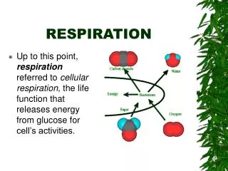
Gas Exchange at Alveolar and Tissue Level • O2 and CO2 Exchange by DIFFUSION • O2 and CO2diffuse down their concentration (partial pressure) gradients • In the inspired air: PO2 = 159mmHg PCO2 = 0.03mmHg • In the alveolar air.: PO2 = 105mmHg PCO2 = 40mmHg • In the systemic capil.: PO2 = 40mmHg arterial end PCO2 = 46mmHg • This is called oxygen cascade (incremental drops in the PO2 from the atmosphere to the mitochondria).
Things that Reduce O2 Exchange • Barometric Pressure • Initial Inspired Fraction of O2 • Humidification (before and after) • Alveolar Mixing • Diffusion Limits • Mixing with Deoxygenated Blood • Extraction by Tissue
Things that Reduce CO2 Exchange • Rate of Production of CO2 • Total Buffer of CO2 • Diffusion (not very limited) • Alveolar Mixing
Which of the following changes occurs during strenuous exercise? • (A) Ventilation rate and O2 consumption increase to the same extent • (B) Systemic arterial PO2 decreases to about 70 mm Hg • (C) Systemic arterial PCO2 increases to about 60 mm Hg • (D) Systemic venous PCO2 decreases to about 20 mm Hg • (E) Pulmonary blood flow decreases at the expense of systemic blood flow
Diffusion Across the Alveolocapillary Membrane • Gases diffuse from the alveoli to the blood in the pulmonary capillaries or vice versa across the thin alveolocapillary membrane • Which made up of: • the pulmonary epithelium • the capillary endothelium • their fused basement membranes
Henry’s Law • When a liquid and a gas, such as blood and alveolar air, are at equilibrium, the amount of gas dissolved in the fluid reaches a maximum value. • According to Henry’s law, this value depends on: • Partial Pressure of the gas • Temperature of the fluid. • Solubility of the gas in the fluid
Rate of Diffusion of Gases through Fluids • affected by: • Partial pressure gradient of gases • Solubility of gas (CO2 is 20X more soluble than O2 in body tissues and can diffuse 20X more rapidly). • Temperature of solution (more gas can be dissolved in cold water than in warm water)
Rate of Diffusion of Gases through Fluids • Surface area of alveolar membrane (N = 50-100 m2, constant) butcan change under abnormal conditions such asemphysema. • Thickness of barrier separating air and blood across alveolar membrane (N = 0.2 - 0.6µm, constant) but can change under abnormal conditions such as pulmonary edema and pneumonia.
Rate of Diffusion (exchange) • Fick’s law of diffusion: the amount of gas diffusing per unit time isinverselyproportional to the thickness of the barrier anddirectlyproportional to the surface area of the barrier. • Where, • D = diffusion rate • SA = surface area • DC = diffusion coefficient • PD = pressure difference D = SA x DC x PD Thickness
Diffusing Capacity of Lung • Amount of gas that diffuses through respiratory membrane per minute per mmHg pressure gradient • Diffusing capacity for O2at rest, is about 25 mL/min/mm Hg. • Diffusing capacity for O2increases to 65 mL/min/mm Hgor more during exercise. • Diffusing capacity for O2reduced in diseases that cause fibrosis of the alveolar walls.
Diffusing Capacity of Lung • Diffusing capacity for CO2 = 20 x that of O2 • Diffusing capacity for CO2 during rest is 400 - 450 ml/minute/mmHg • Diffusing capacity for CO2 increases to 1200 - 1300 ml/minute/mmHg during exercise
Which person would be expected to havethe largest A–a gradient? • (A) Person with pulmonary fibrosis • (B) Person who is hypoventilating due to morphine overdose • (C) Person at 12,000 feet above sea level • (D) Person with normal lungs breathing 50% O2 • (E) Person with normal lungs breathing 100% O2