Download

1 / 77
770 likes | 903 Views
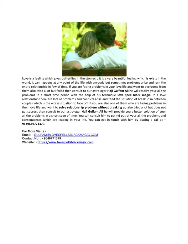
BPPV is the most common cause of vertigo. Read more about vertigo symptoms, get to know what is vertigo and it's treatment and how to cure it with migraine doctor.<br><br>

E N D
VERTIGO – MAKING IT SIMPLE DR.ANITA BHANDARI CONSULTANT NEUROTOLOGIST VERTIGO AND EAR CLINIC, JAIPUR
MATTER OF CONCERN More amenable to treatment -more sinister consequences
Seconds – late ototoxicity Minutes – BPPV, TIA Hours – Meniere’s disease , Migraine related vertigo Days – Vestibular neuritis Months – years - Hysterical
EQUILIBRIUM Spatial orientation Ocular stabilization Postural control
NEUROTOLOGICAL EVALUATION A battery of tests Many systems to be evaluated to assess structural and functional integrity
For An ENT Specialist, We look at the ears first. In vertigo --> eyes are most important
SVV Otoliths act as gravito-inertial force detectors SVV is a psychophysical measure of the angle between perceptual vertical and true/gravitational vertical Also used to measure vestibular rehabilitation Compensated utricular hypofunction may be detected on dynamic SVV testing. The defect will be unmasked on eccentric rotation because any otolith function asymmetry will be enhanced.
SUBJECTIVE VISUAL VERTICAL AND HORIZONTAL Pt is asked to adjust the orientation of a luminous bar until they perceive it as vertical SVV – saccule and its central pathways SVH – utricle and its central pathways Pinar et al reported changes in SVV and SVH in >25% pts of chronic dizziness concluding that evaluation of the otolith system is mandatory
SVV FINDING CONDITION Normal range Upto 2° deviation Ipsiversive tilt – >2o peripheral vestibular disorder pontomedullary lesion thalamic lesion Controversive Pontomesencephalic lesion parietoinsular vest. lesion Migraine Abnormal, little literature
CRANIOCORPOGRAPHY Developed by Claussen [1968] Assessment of vestibulospinal system Photographic recording of head and body movement during gait testing Evaluation includes Romberg, Tandem walking and Unterburger’s test
CCG : PROCEDURE Done in dark room Pt is blindfolded Pt wears a helmet with LED lights Path of the pt is recorded using an SLR camera Result depends on vestibular system only as visuals cues cut off – pt is blindfolded and by stepping in one place, the soles intermittently lose contact with the floor thus reducing somatosensory input
NORMAL PARAMETER OF CCG [CLAUSSEN] PARAMETER NORMAL RANGE- LOWER BORDER NORMAL RANGE- UPPER BORDER Longitudinal displacement 30.03 cm 113.35 cm Lateral sway 5.17 cm 16.15 cm Angular deviation 55.13° (right) 48.37° (left) Body spin 82.21° (right) 82.89° (left)
INTERPRETATIONS OF CCG PATHOLOGY CCG FINDINGS Peripheral vestibular lesions Ipsilateral deviation Brainstem lesion, bilateral peripheral vestibulopathy Enlarged lateral sway, no angular deviation CPA tumors, PICA synd. Contralateral deviation, enlarged sway
ANGULAR DEVIATION TO LEFT
ANGULAR DEVIATION TO LEFT
HEAD IMPULSE TESTING Introduced by Halmagyi and Curthoy Simple, fast, reliable Tests scc function – can evaluate all 3 pairs Measures high freq. vestibular response in 3 dimensions
HEAD IMPULSE TEST VHIT – using Video Frenzel glasses Test for gaze stabilization during rapid translation of head Assesses the peripheral utricular system and superior vestibular N A corrective saccade after VHIT indicates hypofunction of same side
HIT : PROCEDURE Subject seated upright with eyes focused on an fixed object Unpredictable , low amplitude [10 – 20°] head rotation with high acceleration Angular VOR generates compensatory eye movements equal in amplitude and opposite in direction to stabilize gaze
HEAD SHAKING TEST Nystagmus indicates an imbalance in vestibular tone between the 2 sides Not seen in bilateral vestibular dysfunction
HEAD SHAKING – DOWN BEATING NYSTAGMUS
DYNAMIC VISUAL ACUITY TEST Functional test of VOR Comparison of visual acuity with head still to VA with head moving Reduction by 2 lines indicates dysfunction of VOR as seen in bilateral peripheral vestibulopathy Improvement with rehab will improve DVA Early sign of vestibular toxicity
BPPV AND PARTICLE REPOSITIONING MANEUVERS
The ampulla contains the cupula – a gelatinous mass with the same density as the endolymph.Cupula forms an impermeable barrier across the lumen of the ampulla. Hence the particles in scc may only exit via the end with no ampulla.
POSTERIOR CANAL BPPV POSTERIOR CANAL BPPV Most common– posterior canal is most gravity dependent in upright and supine position Once debris enter the post. canal ,the cupula at the shorter most dependent arm trap the debris. Debris can exit only through the longer arm through the crus commune [non-ampullary]
DIX-HALLPIKE MANEUVRE
EPLEY EPLEY’ ’S MANEUVER S MANEUVER
SEMONT SEMONT’ ’S MANEUVER S MANEUVER Liberatory maneuver for pBPPV and cupulolithiasis Used to overcome otoconia jam after Epley’s maneuver
SEMONT SEMONT’ ’S MANEUVRE S MANEUVRE
BRANDT – DAROFF EXERCISES BRANDT – DAROFF EXERCISES Used as a home program Indications o Posterior canal cupulolithiasis o Persistant posterior canal canalithiasis Mechanism o Dislodge debris attached to cupula o Habituation through central compensation
BRANDT-DAROFF EXERCISES
BRANDT – DAROFF EXERCISES BRANDT – DAROFF EXERCISES Things to remember o The exercises may dislodge more otoconia from the utricle causing an increase in symptoms. o May cause multiple canal involvement. o Important to hold for 30 seconds in each position.
HORIZONTAL SCC BPPV HORIZONTAL SCC BPPV Pagnini-McClure maneuvre Geotropic nystagmus – debris are away from ampulla , side showing stronger nystagmus is the side involved Apogeotropic nystagmus – indicates cupulolithiasis
McCLURE PAGNINI MANEUVER McCLURE PAGNINI MANEUVER SUPINE ROLL TEST SUPINE ROLL TEST