Download
1 / 34
360 likes | 567 Views
RasMol workshop. Protein Structure and Function A. http://www.expasy.org/sprot/sprot-top.html. PDB – Protein Data Bank. http://www.rcsb.org/pdb/. http://pdb.tau.ac.il. Protein visualization.

E N D
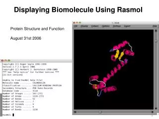
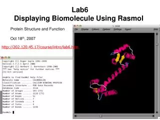
RasMol workshop Protein Structure and Function A
PDB – Protein Data Bank http://www.rcsb.org/pdb/
Protein visualization The structure allows better understanding of the structure-function relationship, and is an important starting point for many kinds of research. Visualization tools (working on PC): RasMol SwissPDBviewer (very powerful and contains many functions) Chime (The same as RasMol but without command line) → work trough the web Protein Explore (more advanced than RasMol)
RasMol- Main Menu פתיחת קובץ אינפורמציה שיש ב -PDB סגירת קובץ
RasMol - Display קווים בין אטומים
RasMol - Display הצגת פחמנים
RasMol - Display יותר משקל לקשרים בין אטומים
RasMol - Display נתוני רדיוס ה –VDW של כל אטום – מיקום במרחב של כל אטום בהתאם
RasMol - Display אלמנטים של מבנה שניוני, קו המחבר בין C-alpha
RasMol - Display מחזק מבנה שניוני, למשל ראש חץ הוא C- טרמינל
RasMol - Color תצוגה מסוימת בו לכל אטום צבע מסוים לכל חומצה אמינית צבע משלה עד במה המקום קבוע במבנה: אדום- חופש גדול, כחול-קבוע מאוד מבנה שניוני – צהוב- beta shith, כחול – loops and turns, אדום – alpha helix, לבן – rendom coil. לקבוע צבע כבר בקובץ ה- PDB
RasMol - Options מוריד 50%, טוב לראות חללים.
RasMol - Options בעזרת משקפיים מיוחדים, אפשר לראות ב-D3.
RasMol - Options סימן כל המולקולה או קטעים נבחרים בשם והמספר של חומצת האמינו.
RasMol - Options לראות או להסתיר מימנים לראות או להסתיר קבוצות לא חלבוניות או ליגנדים נותן עומק לתמונה מקור אור נמצא בפינה כלשהיא של המסך ומקרין אור על המולקולה.
RasMol- Command Line רושמים את הפקודות שאנו רוצים לבצע
The Select Command Select – מגדירה את האזור שעליו נפעל במולקולה - סט האטומים שיעבור מודיפיקציות ע"י שרשרת הפקודות הבאות. הפרמטר של פקודת ה- select הוא “atom expression”. ה - atom expression מגדיר באופן ייחודי קבוצה שרירותית של אטומים בתוך מולקולה. הוא יכול להיות גם: Primitive expressions, Predefined sets, Comparison operators, Within expressions, or logical combination of all above mentioned.
The primitive expressions allow to select by: • Atom number - select atomno=102 • Residue – select Val52 (select resno=52) • Chain id – select :a • List of residue numbers – select 14,92, 46 • Range of atom numbers – select atomno=>35 • A wildcard can be used to specify a whole field: • * Any number of characters Atom or residue type – select *.sg (this will select all Sulphur atoms in Cistein’s side chain) • ? Single character wildcard – select ser70.c? – will select all carbons in all • serine residues.
The within expressions defines the neighbors of a given set of atoms: select within (4.0, backbone) Distance: the cut-off in Å Where containing decimal point Set of atoms Example : all atoms not further than 3.5Å from Ala35: Select within (3.5, Ala55)
The predefined sets are groups of atoms given the definite names: select helix select hoh (water molecules) select protein There is a list with the predefined sets In order to display only what we selected, use the command: restrict selected
Boolean Expressions And – המשותף לשני תנאים Or – חיבור בין שני תנאים (גם את זה וגם את זה) Not – מה לא לכלול דוגמאות: select tyr and :a → all tyr in ‘a’ chain select tyr or :a → all tyr in the molecule and all ‘a’ chain select not (try,:a) → all the molecule beside try and ‘a’ chain
Exercise • Load the 1GCD.pdb (file → open) • Go over the display menu and try all of the options • Set the display on wireframe and try the color menu • Set the display on cartons and try the color menu again • Than, try the command line: • ribbons • wireframe 40 • spacefill 120 • spacefill off • select Ser • spacefill 150 • color cpk • zoom 200
select all wireframe 40 (If it doesn't work, do Display => wireframe in the menu) color chain hbonds on (how much Hbonds are their?) hbonds 30 color hbonds green hbonds off select hetero and not hoh spacefill 120 color CPK (Touch the selected atoms with the mouse and look on the command line) select water spacefill 120 color magenta select ligand spacefill 300
select asp102,his57,ser195 or ligand restrict selected center selected color CPK Display => balls and sticks (on the menu) labels on labels off (try also the option menu- labels, what is the difference?) Select ligand Display sticks set picking distance (pick a pair of atoms which you want to know their distance) set picking distance off
Which atoms are present besides the protein? Show only them. Display each hetero atom type in different presentation. • Show only the ligand (inhibitor) and the oxyanion pocket (gly193,ser195) • Color them in CPK. Display the inhibitor in sticks and the protein’s oxyanion pocket in balls and sticks. • Label the residues and the inhibitor (not every atom, just the number and type) • Measure the distance between the gly’s nitrogen and the ligand’s oxygen.
Select all the protein. How many secondary structures does the protein contains? • Show only the helixes, center them on the screen. • Select the resides that are within the radius of 8.0 Å from the inhibitor. Display only them and the inhibitor. Color the inhibitor in CPK. Color the hydrophobic residues in blue and charged residues in magenta (the others in white). What do you see?
Carboxy peptidase exercise • חפשו ב-PDB את המבנה של הקרבוקסי פפטידז ותעלו את הקורדינטות שלו ב-RasMol. • הסתכלו ב-PDB, מהי השיטה בה קבעו את המבנה? מה זה אומר לנו? • בעזרת ה-PDB, זהו את האתר הפעיל. האם יש ליגנד? מיהו? מה תפקידו? האם יש יותר מליגנד אחד? • 2. זהה בעזרת RasMol את הליגנדות. • 1. הראה את הקשרים הקורדינטיבים של האבץ (רמז ← קשר קורדינטבי הוא בין Å2-3). • מדוד את המרחקים של הקשרים הקורדינטיבים. • 2. זהה את המעכב והראה את הקשרים שהוא יוצר בעזרת RasMol (רמז ← השתמש ב- PDB Sum).
Select hetero (or not protein) Restrict selected Select hoh Display wireframe Select within (3.0, ZN) Display balls and sticks Select 196,69,72 Display balls and sticks Option labels Select within (5.0,cpm) Restrict selected Display balls and sticks Select cpm Display spacefill
Select 145 Display spacefill Display balls and sticks Select 248,145 Display spacefill Display balls and sticks Select 270,196 Display spacefill Display balls and sticks Select 127 Display spacefill Display balls and sticks