Download

1 / 96
1.29k likes | 4.52k Views
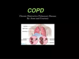
Pathology lecture for medical students. Pathologenesis of COPD,

E N D
“Within the mind are all the resources required for successful living. Ideas are present in the consciousness, which when released and given scope to grow and take shape, lead to successful events” - Wings of Fire: An Autobiography of Dr. APJ Abdul Kalam.
“ Whether you think you can or you can't, you are right…!” – Henry Ford
CPC14: Mrs. PT. 64y Fem. SOB Mrs. P.T. 64 y female, to ED at TTH. Worsening SOB, years, worst today - At rest • Previously SOB after exertion- COPD. • Gradually worsening over the years, now O2 at night. • Sputum chronic clear white, but light brown today. • Cough - on and off for years, No wheeze, no hemoptysis, • Chest pain worse with breathing today, fever, sweaty. • Smoked 3 packs/day for 35 years; quit 7y ago. Foreign travel: Norfolk Island 3wks. No health problem. RX: Pneumonia, COPD, Emphysema, CCF
CPC: 2011 – 58M Chronic cough. Trevor is 58 year old Caucasian man, years of coughing. pneumonia 3 years ago; bronchitis several times a year, Dyspnoea, Hoover’s & Campbell sign positive, leans forward for breathing. heavy smoker (30+/day); quit 3 years ago. Previous PFT: FEV1 = 1.3 FVC = 2.6 FEV1/FVC = 50%. (too sick to perform PFT today.) • What is the significance of… 1. Leaning forward – “arms on knees” 2. Intercostal in-drawing 3. Hoover's sign ? Tracheal tug ? 4. Campbell's sign ? 1. 2. 3. 4. Pathogenesis of symptoms ? Further questions ? Differential diagnosis ? Learning issues? PFT
Clinical Feature - ? Pathogenesis SOB, Dyspnoea Cough Wheezing, Stridor. Rhonchi, Crackles/ Rales. Dullness Hyper-resonance Hyperventilation Tachypnoea Pleural rub Pleurisy (pleuritis) Sleep apnoea central/obstructive. • White sputum (frothy) • Rusty, Yellow, green sputum • Haemoptysis • Night sweats • Sleep apnoea • Kussmaul’s breathing. • Ataxic breathing • Apneustic breathing. • Cheyne-Stokes breathing. • Paradoxical breathing • Resp acidosis / alkalosis.
COPD: Questions What is Chronic Bronchitis & Emphysema ? Pathogenesis of COPD/CB/Emphysema ? Smoking – Disease, Pathogenesis ? Difference.. Obstructive / Restrictive disease ? What findings on PFT expected ? How is he maintaining normal pO2 & pCo2 ? why is he pink & puffing ? What Gross & Microscopic features in his lung ? What complications are expected ? Pneumoconiosis, TB, DPF,
COPD: Questions Bronchiectasis ? Atelectasis ? Lung abscess ? Honeycomb lung ? Pneumothorax ? Tension pneumothorax? Pleural effusion Emphysema types. • Panacinar - α1AT def. • Paraseptal • Irregular • Bullous • compensatory Tuberculosis Primary, secondary, anergic, milliary, caseous, fibrocaseous, fibrosing. Asthma – types. Atopic, non-atopic, drugs, occupational etc. Pathogenesis, gross, micro Curschmann spirals, Charcot- Leyden crystals. Pneumoconioses Silicosis, Anthracosis, Asbestosis IPF – Ideopathic pulm. fibrosis. • • • • • • • • • • Text Book: Robbins Basic Pathology, 8th or 9th edition
Core Learning Issues: Major: • COPD Pathology – Restrictive / Obstructive. • Emphysema & Bronchitis – types, pathology & clinical. • Pathology of Tobacco related disorders. • PFT - FEV1/FVC & its use and interpretation. • Pneumonia – types & Pathology (Lobar, Broncho & Int.) • Allergic bronchitis, Asthma, Tuberculosis * self study. Minor: • Pneumoconiosis – Asbestosis, silicosis etc. • Cor pulmonale, Pulmonary hypertension. • Diffuse Pulmonary Fibrosis & honeycomb lung. • Acute lung injury - ARDS, DAD, HMD of infants. • Sarcoidosis ?
Normal Lung Tr.Air Sh Br. Mar. Colon air
Lung Histology Bronchiole Alveoli Type 2 Type 1-Pneumocyte
There is only one secret to staying young, being happy, and achieving success - You've got to “enjoy what you do”…! Including studying pathology…..
Pathology of COPD, Chronic lung diseases & Pneumonia* Dr. Shashidhar Venkatesh Murthy. A/Prof. & Head of Pathology
Percent Change in Age-Adjusted Death Rates, U.S., 1965-1998 4thleading cause of death, women more, smoking >80% Proportion of 1965 Rate 3.0 Coronary Heart Disease 3.0 All Other Causes Stroke Other CVD COPD 2.5 2.5 2.0 2.0 1.5 1.5 1.0 1.0 0.5 0.5 –59% –64% –35% +163% –7% 0.0 0 1965 - 1998 1965 - 1998 1965 - 1998 1965 - 1998 1965 - 1998
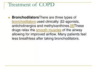
Pathology of Smoking Research evidence: smoking disease >4000 chemicals, 43 carcinogens. >90% of COPD is due to smoking. 15% of smokers develop COPD. Injury & inflammation central to damage. Smoking a single cigarette results in an acute increase in neutrophils at 1 hour. Increased neutrophils in smokers, which decrease following reduction/quitting. Patients with COPD have sputum neutrophilia that persists even after cessation of smoking.
Smoking related diseases: Non-neoplastic: • Bronchitis, pneumonia, bronchiectasis. • Chronic bronchitis, emphysema (COPD) • Atherosclerosis IHD, Stroke, MI. • Gastritis, Peptic Ulcer, Oesophagitis. • Arteriosclerosis – Berger’s disease. Neoplastic: • Lung Cancer (many types) • Oral, laryngeal & oesophageal cancer. • Carcinoma of bladder, Pancreas, cervix, larynx.
Ask, Aspire, Achieve..! …..Ask questions J. Robin Warren Australian 2005 Nobel price in Medicine - at RCPA conference 2006 Sydney.
Pathogenesis of COPD Dr. Shashidhar Venkatesh Murthy. A/Prof. & Head of Pathology
Pathogenesis of COPD 1. Smoke, irritants, carcinogens. 2. Tissue irritation & destruction 3. Inflam Mucous production. 4. Airway damage Narrowing. 5. Alveolar damage widening. Increase in • Alveolar marcrophages • CD8 Lymphocytes • Neutrophils • Proteases. Airway inflam Bronchitis Alveoli damage Emphysema. Both COPD. α1AT def.. Emphysema Bronchitis Emphysema
The mind is everything. What you think you become! -- Buddha
Pathogenesis – Chronic Bronchitis Irritation COPD Initiation Promotion Ca. Chronic Bronchitis K-Ras C-myc 3p p53 Pathogenesis: 1. Smoking – carcinogens 2. 3p – tumor suppressor gene loss 3. Mutations (p53, KRAS, EGFR..) 4. Dysplasia 5. Infiltration 6. Spread 7. Metastases.
Chronic Bronchitis - Emphysema: Predominant Bronchitis 40-45 Mild; late Early; copious sputum Common Repeated Predominant Emphysema 50-75 Severe; early Late; scanty sputum Occasional Terminal Age (yr) Dyspnea Cough Infections Respiratory insufficiency Cor pulmonale Airway resistance Elastic recoil Chest radiograph Appearance Common Increased Rare; terminal Normal or slightly increased Low Hyperinflation; small heart Normal Prominent vessels; large heart Blue bloater Pink puffer
“Know more today about the world than yesterday and lessen the suffering of others. You'd be surprised how far that gets you.” ― Neil deGrasse Tyson
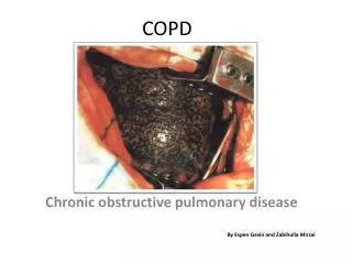
Smokers Lung: COPD - Emphysema Upper lobe – black spots of anthracotic (Co2) pigmentation (centre of each lobule). Centrilobular alveolar destruction with carbon pigmentation. Other types: * • Panacinar • Paraseptal • Bullous • Interstitial • Irregular.
Normal - Emphysema
Normal Emphysema
Normal - COPD CB Emphysema
Centrilobular Emphysema: Pink Puffer: Lean/weight loss No cyanosis Forward stooping Barrel chest Flat diaphragm Hyperlucent Lung
Whether you think that you can or that you can't, you are right…! – Henry Ford
Chronic Bronchitis: Normal Chronic Bronchitis • Squamous Metaplasia. • mucous gland. Hyperplasia
Emphysema: Other types Panacinar • Congenital, α1AT def. Paraseptal, Bullous • Old scar. Irregular • Past diffuse scaring.
Complications of COPD: 1. Pneumothorax, Infections, Bronchectasis. 2. Polycythemia – hypoxia. 3. End-stage lung disease. 4. Acute Exacerbations. 5. Cor Pulmonale – Right heart failure. • syncope, hypoxia, pedal edema, passive hepatic congestion and death. Lung Cancer LV RV
Bronchiectasis: Permanent dilatation & Infection of bronchi. Cough, copious purulent sputum, mixed infections. Lower lobes common Secondary to COPD, Pneumonia or localised obstructions. Complications: • Pneumonia, empyema, septicemia, meningitis. Types: • Cylindrical, Saccular, Fusiform (no
Bronchiectasis: • Permanent dilatation with secondary infection • Pus* filled, *visible bronchi till *periphery. • Abundant, greenish, sputum, mix culture +ve.
COPD Summary: Definition: Progressive Irreversible Chronic airflow limitation due to inflammatory response to noxious substances. Progressive decrease in PF. Diagnosis: FEV1<80%, FEV1/FVC <70% Etiology • >90% smoking (15% of smokers COPD) • Pollution, 1AT def. , genetic susceptibility, Asthma, idiopathic. Clinical Syndromes: CB, Emphysema, COPD, Asthma, & Bronchiectasis. Complications: Pulmonary failure, RS Cardiac failure, Endstage disease, Cancer.
Self Study - Restrictive Lung Disorders. - IPF, Diffuse fibrosis, - Honeycomb lung. - Pneumoconiosis - Silicosis, Asbestosis, Coal. -Tuberculosis. - Asthma - Good pasteur’s syndrome. - DAD: ARDS/HMD
Pneumonia: Clinical vignette 50y man, alcoholic, high fever, cough, copious foul smelling brown sputum, pleuritic rt sided chest pain. HPC: wife reports that he was brought home in a semi- conscious state a few days ago – drunk. Thin, distressed, pursed lip breathing, using accessory muscles of respiration, cannot speak in full sentences, leaning forwards…* 39°C, RR-22/min, peripheral cyanosis. Chest-rib in-drawing, diminished air entry, soft expiratory wheezes, bronchial breathing L lower post chest. PE: fever, consolidation right middle and lower lobes. sputum microscopy - abundant PMN and mixed oral flora. Pathogenesis, Differential diagnosis…..?
Pneumonia: Questions What is pneumonia? Types? pathogenesis? Lobar, Broncho & Interstial pneumonia? Community acquired / Nosocomial / hospital acquired pneumonia ? What is the difference ? Acute, Chronic & recurrent pneumonia? Typical, Atypical pneumonia? Common organisms causing pneumonia? Microbiology – lab diagnosis, culture, tests. Gross and microscopy of pneumonia. Phases of pneumonia – Congestion, Red hepatization, Grey hepatization, Resolution? Complications of pneumonia? Lipoid pneumonia, Carcinomatous & Aspiration pneumonia ?
Goodpasture Synrome: ?Etiology ?Clinical ?Pathogenesis ?Diagnosis.
Diffuse Alveolar Damage (DAD): ?Etiology ?Clinical ?Pathogenesis ?Diagnosis. Diffuse bilateral consolidation with honeycombing in both lower lobes, focal in upper lobes. Microscopy: Hyaline membrane in ARDS.
Saddle Thomboemobulus: ?Etiology ?Clinical ?Pathogenesis ?Diagnosis.
Clinical Problem Solving - Course Free online course from UCSF https://class.coursera.org/clinprobsolv-001/class/index