Download
1 / 1
10 likes | 136 Views
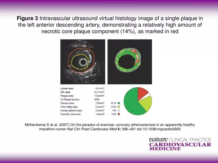
Figure 3 Intravascular ultrasound virtual histology image of a single plaque in the left anterior descending artery, demonstrating a relatively high amount of necrotic core plaque component (14%), as marked in red.

E N D
Figure 3 Intravascular ultrasound virtual histology image of a single plaque in the left anterior descending artery, demonstrating a relatively high amount of necrotic core plaque component (14%), as marked in red Möhlenkamp S et al.(2007) On the paradox of exercise: coronary atherosclerosis in an apparently healthy marathon runner Nat Clin Pract Cardiovasc Med4: 396–401 doi:10.1038/ncpcardio0926