Download
1 / 46
460 likes | 655 Views
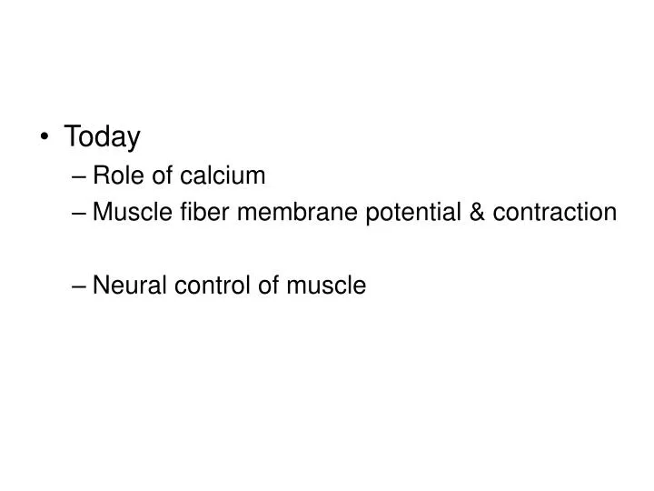
Today Role of calcium Muscle fiber membrane potential & contraction Neural control of muscle. Role of calcium. Tropomyosin. Troponin complex. Troponin and Tropomyosin bind to actin block the actin – myosin binding sites Troponin is a calcium binding protein.

E N D
Today • Role of calcium • Muscle fiber membrane potential & contraction • Neural control of muscle
Role of calcium Tropomyosin Troponin complex • Troponin and Tropomyosin bind to actin • block the actin – myosin binding sites • Troponin is a calcium binding protein
When Troponin binds calcium it moves Tropomyosin away from the actin-myosin binding site Ca Ca
Where does Calcium come from? • Intracellular storage called Sarcoplasmic Reticulum • Surround each myofibril of the whole muscle • Contains high concentration of calcium • Transverse Tubules connects plasma membrane to deep inside muscle
Myofibril Transverse tubules Sarcoplasmic Reticulum Transverse tubules
So far: • Actin and myosin will bind to each other • Troponin / tropomyosin inhibit this • Calcium removes inhibition
What controls muscle calcium? • What else do we know? • Neurons initiate muscle contraction at NMJs by generating postsynaptic potentials (some muscle fibers have APs) • Maybe muscle membrane potential is important
Excitation-Contraction coupling:1 Stimulate nerve Vm Tension Force Transducer Muscle fiber ‘twitch’ Muscle AP Tension Vm Time
Excitation-Contraction coupling:2 Vm Tension Muscle fiber Force Transducer Vary [K+] outside 1.0 Conclusion: Muscle contraction occurs with Vm depolarization Tension 0 -70 -60 -50 -40 -30 Vm (mV)
Why T-tubules important? Stimulate near T-tubule see contraction of adjoining sarcomeres No contractions ‘Local stimulation’ T-tubule
Membrane depolarization or APs carried deep into the muscle by T-tubules Motor nerve T-tubule + Neurotransmitter receptors SR
Text Fig 10-21 Myofibril Transverse tubules Sarcoplasmic Reticulum Transverse tubules
My SR Ryanodine Receptor Dihydropyridine receptor T-tubule SR myoplasm
Ca++ Ca++ Ca++ SR Ca++ pump Myoplasm (intracellular) _ _ _ + _ + _ + + _ + + _ _ + _ + _ T-tubule (extracellular) _ + + + _ +
Summary of events • Synaptic Depolarization of the plasma membrane is carried into the muscle by T-Tubules • Conformational change of dihydropyridine receptor directly opens the ryanodine receptor calcium channel • Calcium flows into myoplasm where it binds troponin • Calcium pumped back into SR
Neural Control of Muscle • Voluntary • Reflex
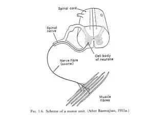
Neural control of muscle contraction Motor Pool: all of the motor neurons that innervate a single muscle Motor Unit: single motor neuron and all the muscle fibers it innervates • a few fibers 1000s of fibers
Size of the motor units determines precision of movement • Fingers have small motor units, legs have big motor units • Recruitment of twitch fibers • Smallest motor units to a single muscle are recruited first • Why? Allow smooth generation of movement
Third Second First Individual myofibrils Motor neurons Whole muscle Little force More force Even more force 1+2+3 = maximum force
Reflex control of muscle contraction Two sensory receptors • Muscle Spindle • Monitors muscle length • Golgi Tendon Organ • Monitors muscle tension
Muscle Spindle Group I and II Sensory fibers • Motor neurons Muscle Spindle Intrafusal Muscle fibers Extrafusal Muscle fibers • Motor neurons
Muscle spindle nerve Extrafusal muscle fibers
motor neurons innervate extrafusal muscle fibers and cause the muscle to contract • motot neurons innervate only the intrafusal muscle fibers and cause them to contract • The sensory endings in the muscle spindle are activated by muscle lengthening
Muscle length Isolated muscle Spinal cord Ia sensory neuron AP Muscle stretch AP APs in sensory • Motor neurons APs in motor Longer
Effect of muscle spindle • When muscle stretches, spindle stretches • Increase APs in 1a sensory neuron • Increase APs in motor neuron • Muscle contracts and returns to original length (almost)
When muscle contracts, spindle shortens • Might expect activity of spindle to decrease • BUT • To maintain sensitivity of the spindle, the intrafusal fibers also contract • Controlled by motor neurons
Muscle Spindle Group I and II Sensory fibers • Motor neurons • Motor neurons Extrafusal Muscle fibers Intrafusal Muscle fibers
Motor neuron Ia sensory neuron APs in sensory - Motor neuron only stimulate shorter Muscle length record longer
Motor neuron Ia sensory neuron APs in sensory - Motor neuron only stimulate shorter Muscle length record longer Motor neuron APs in sensory - and Motor neurons
Muscle Spindle motor neurons • permit muscle spindle to function at all muscle lengths • Maintains sensitivity of the spindle
Spinal cord Inhibitoryinterneuron Ia sensory neuron Motor neurons Muscle spindle
Golgi Tendon Organ • Operates like muscle spindle, but monitors muscle tension (force) • Negative feedback because they inhibit the muscle they are located in
Golgi Tendon Organ Very little at rest Increased APs during contraction APs from GTO shorter Muscle length longer
Spinal cord Inhibitoryinterneuron sensory neuron Motor neurons Golgi tendon organ
Muscle Spindle Response Tendon Organ Response Decrease APs Passive Stretch Increase APs Active Contraction Increase APs No change
Summary • Muscle Spindles • Monitor muscle length • When activated cause contraction • Golgi Tendon Organ • Monitor muscle tension • When activated reduce contraction
Whole muscle physiology • Types of skeletal muscle fibers • Neural control of muscle contraction • Production of force
Classification of muscle fiber types • Electrical properties of muscle membrane – does muscle have APs? • Maximal rate of contraction (Vmax) • determined by myosin ATPase activity • Density of SR calcium pumps • Density of mitochondria and blood supply
Vertebrate Skeletal Muscle Fiber Types: • Tonic • Twitch (or Phasic) • Slow oxidative (Type I) • Fast oxidative (Type IIa) • Fast glycolytic (Type IIb)
Tonic fibers • Very slow contractions • Do not produce APs do not twitch • Postural muscles
Twitch muscles • Slow oxidative (Type I) • Contract slowly • Resist fatigue • Postural • Fast Oxidative • High rate of contraction • Moderately resistant to fatigue • Rapid, repetitive motion (flight muscles migratory birds) • Fast Glycolytic • Rapid contraction • Rapid fatigue
Non-twitch fibers • Many arthropods (crayfish, insects) do not have muscle APs • Rather they have graded synaptic potentials • Calcium released from SR in graded manner • Degree of contraction depends on summation and facilitation of neural input
Muscle AP Tension Vm Time Summation of Synaptic Potential Tension Vm Time
Non-twitch fibers • Graded potential graded contraction • Even large motor units can have precise contraction