Download
1 / 25
300 likes | 631 Views
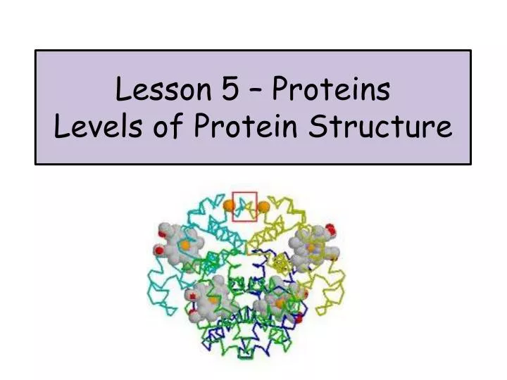
Lesson 5 – Proteins Levels of Protein Structure. Primary 1º Structure. The primary structure is simply the sequence of amino acids in a protein. Chains of amino acids are written from the amino terminus (N-terminus) to the carboxyl terminus (C-terminus).

E N D
Primary 1º Structure The primary structure is simply the sequence of amino acids in a protein. Chains of amino acids are written from the amino terminus (N-terminus) to the carboxyl terminus (C-terminus).
Usually the amino acids are represented by their 3 letter abbreviations. Less commonly you will see the full name of each amino acid or even the single letter designations.
Secondary 2º Structure There are 2 types of secondary structure α-helix and β–sheet.
α-helix The helix is right handed with 4 residues (amino acids) per turn. Hydrogen bonds between oxygen (the C=O group) and hydrogen (N-H group) stabilise the helix.
β-Sheet Polypeptide chains are linked together in a side by side formation with hydrogen bonds. Hydrogen bonds
β-Sheet β-sheets can be parallel or antiparallel.
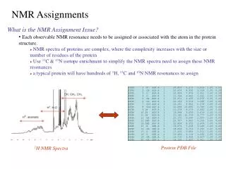
Tertiary 3º Structure The tertiary structure is all about the interactions between the R groups. This involves the folding of the polypeptide chains to give a more complex 3D structure. The complexity of the tertiary structure of proteins has been determined using techniques such as X ray crystallography and NMR (nuclear magnetic resonance).
Hydrophobic Interactions The non-polar R groups (remember – these are hydrophobic) tend to get placed in the centre of the molecule.
Disulphide Bonds Disulphide bonds are strong covalent bonds can form between - SH groups that are found in different parts of the polypeptide. Think? Which amino acid residue contains an –SH group? Cysteine!
Prosthetic Groups Some proteins have associated non-protein groups. These are known as prosthetic groups.
Myoglobin Myoglobin has m –helices linked together with nonhelical sections. It contains a haeme group that is protected in a hydrophobic ‘pocket’.
Stability of Tertiary Structure Protein structures are secured by weak hydrogen bonds and a few disulphide bonds. Despite this they are surprisingly stable structures. This due in the most part to the evolution of proteins that have useful yet stable conformations. Proteins fold to give a structure with the lowest free energy – this will therefore change if the chemical environment changes.
Quaternary 4° Structure Some proteins are made up of 2 or more polypeptide subunits. Haemoglobin is an example of a protein with a quaternary structure – it is tetrameric (made up of 4 parts). It has 2 α–helices and 2 β–sheets. Each of the polypeptide chains has a haeme group and the 4 chains are held together by hydrogen and ionic bonds.
Motifs and Domains When describing proteins we can identify motifs and domains in the protein. Motif – a particular form of secondary structure. An examples include βαβmotif or βbarrel. Domains – regions of a polypeptide chain that fold independently to give distinct regions with potentially different roles
Domain “A protein domain is a conserved part of a given protein sequence and structure that can evolve, function, and exist independently of the rest of the protein chain. Each domain forms a compact three-dimensional structure and often can be independently stable and folded.” Wikipedia Pyruvate kinase has 3 different domains.
Classes and Functions of Proteins Page 38 in your monograph details 9 different classes of proteins. Your task: Prepare a powerpoint, poster or mindmap to include all of the information found on page 38. Try to come up with ways of remembering the functions and some examples of each. There will be a quiz on all of section 2.3 Proteins on Monday November 26th.
Your Task- Proteins Past Paper Questions You will need to access these in the department. Please DO NOT TAKE past papers home – we have limited numbers. 2002 MC Q5 2004 MC Q12 2006 Section B Q1 2008 MC 5 2009 Section B Q6 2. Complete Scholar activities on amino acids. 3. Read and make notes on pages 30-35 Make sure your glossary is up to date. Pg 38 protein task. DUE Monday (November 26th)