Download
1 / 36
390 likes | 986 Views
Post Partum Hemorrhage. Akmal Abbasi, M.D. Post Partum Hemorrhage. Obstetric Haemorrhage: Ranks as the First cause of maternal mortality accounting for 25 – 50 % of maternal deaths.

E N D
Post Partum Hemorrhage Akmal Abbasi, M.D.
Post Partum Hemorrhage • Obstetric Haemorrhage:Ranks as the First cause of maternal mortality accounting for 25 – 50 % of maternal deaths. • Post Partum Hemorrhage though preventable, accounts for the majority of the cases of obstetric hemorrhage, the other causes being – antepartum hemorrhage, abortion, ectopic pregnancy and ruptured uterus.
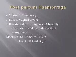
HEMORRHAGE IN THE PREGNANT PATIENT • The normal pregnant patient frequently loses 500 ml of blood at the time of vaginal delivery and 1,000 ml at the time of cesarean delivery. • Appreciably more blood can be lost without clinical evidence of a volume deficit as a result of the 40 percent expansion in blood volume that occurs by the 30th week of pregnancy.
HEMORRHAGE IN THE PREGNANT PATIENT • To understand why the pregnant patient does not exhibit early signs of volume loss, it is important to understand the normal physiologic responses to hemorrhage. • When 1,000 ml is rapidly removed from the circulatory blood volume, vasoconstriction occurs in both the arterial and venous compartments in order to preserve essential body organ flow. • When intravascular volume is lost, blood pressure is initially maintained by increases in systemic vascular resistance.
HEMORRHAGE IN THE PREGNANT PATIENT • As volume loss exceeds 20 percent, the fall in cardiac output accelerates and blood pressure can no longer be maintained by increases in resistance. • Blood pressure and cardiac output fall in parallel after this point. • If the volume loss has occurred more than 4 hours earlier, significant fluid shifts from the interstitial space into the intravascular space will partially correct the volume deficit.
HEMORRHAGE IN THE PREGNANT PATIENT • This movement of fluid, termed transcapillary refill, can replace as much as 30 percent of lost volume. • In more chronic bleeding states, the final blood volume deficit may amount to as little as 70 percent of the actual blood lost.
Relationships among systemic vascular pressure, cardiac output, and blood pressure in the face of progressive blood volume deficit.
ASSESSMENT OF BLOOD LOSS AFTER DELIVERY • Difficult • Mostly Visual estimation (So, Subjective & Inaccurate) • Underestimation is likely • Clinical picture -Misleading • Our Mothers-Malnourished, Anaemic, Small built, Less blood volume
Etiology • Uterine atony (80%) • Retained Placenta • Trauma to genital tract • Coagulation disorders • Uterine inversion
Etiology of PPH The causes of postpartum hemorrhage can be thought of as the four Ts: • tone, • tissue, • trauma, • thrombin
Etiology of PPH Uterine atony • Multiple gestation, highparity, Full bladder, • Anesthesia (halogeneted) & analgesia • Prolonged, augmentedlabor, or • chorioamnionitis, • tocolytic agents
Etiology of PPH UTERINE INVERSION • Incomplete Inversion- Fundus felt through the Cx • Complete Inversion with placenta accreta attached to the fundus • Mostly iatrogenic due to mismanagement of 3rd stage - strong traction on the cord with a relaxed uterus / adherent placenta
Etiology of PPH Placental abnormalities Congenital Bicornuate uterus Location Placenta previa Attachment Acquired structural Leiomyoma previous surgery Peripartum Uterine inversion, uterine rupture, placental abruption Accreta
Etiology of PPH Lacerations and trauma • Unplanned • Vaginal/cervical tear, • surgical trauma • Planned • Cesarean section, • episiotomy
Etiology of PPH Coagulationdisorders • Acquired • DIC, Sepsis, Severe • PET/ Eclampsia • Amniotic fluid embolism • Hepatitis • heparin Congenital Von Willebrand's disease
PREVENTION • Regular ANC • Correction of anemia • Identification of high risk cases • Delivery in hospital with facility for Emergency Obstetric Care. • Local / Regional anesthesia • ACTIVE MANAGEMENT OF 3RD STAGE OF LABOR • 4th Stage of labor - Observation, Oxytocin
ACTIVE MANAGEMENT OF 3RD STAGE OF LABOR • Oxytocics - Routine use in third stage blood loss by 30-40% • Early cord clamping • Controlled cord traction • Inspection of placenta & lower genital tract
MANAGEMENT OF PPH • TEAM- Obstetrician, Anesthesiologist, Hematologist and Blood Bank • Correction of hypovolemia • Ascertain origin of bleeding • Ensure uterine contraction • Surgical management • Management of special situation
MANAGEMENT OF PPH CORRECTION OF HYPOVOLEMIA • Large bore IV line (two) • Crystalloids (RL) • Monitor Urine output • Whole blood / pack cell
MANAGEMENT OF PPH ENSURE UTERINE CONTRACTION • Palpate fundus • Uterine massage • Bimanual compression • Compression of Aorta against sacral promontory • Foleys catheters
MANAGEMENT OF PPH OXYTOCICS • Oxytocin: • Ergometrine • Prostaglandins
MANAGEMENT OF PPH SURGICAL TREATMENT Depends on • Extent & cause of hemorrhage • General condition of patient • Future reproduction • Experience & skill
MANAGEMENT OF PPH SURGICAL TREATMENT • Repair of trauma if any • Uterine A. ligation • Utero ovarian A. Ligation • Internal Iliac A. Ligation • Brace suturing of Uterus • Hysterectomy • Angiographic embolization
MANAGEMENT OF PPH RETAINED PLACENTA • EUA & Manual Removal • If Placenta accreta- • Observation • Cytotoxic drugs- Methotrexate • Hysterectomy
MANAGEMENT OF PPH ACUTE INVERSION OF UTERUS • Manual replacement- • Under GA / Uterine relaxant • Hydrostatic method • Surgical method ( Usually delayed procedure)
MANAGEMENT OF PPH MANAGEMENT OF DIC • Fresh blood transfusion • Blood products • Cryoprecipitate • Fresh frozen plasma • Platelet concentrate
MORBIDITY & MORTALITY from PPH • Shock & DIC • Renal Failure • Puerperal sepsis • Lactation failure • Blood transfusion reaction • Thromboembolism • Sheehan’s syndrome • >25% Maternal deaths are due to PPH
A, A fetal heart rate tracing at 37 weeks' gestation in a patient involved in an automobile accident in which the maternal abdomen struck the steering wheel. Fetal movements were present, and no periodic changes were observed. Uterine contractions were occurring every 2 to 3 minutes.
B, Eight hours later, repetitive late decelerations are now present. An asphyxiated fetus was delivered, and evidence of grade 3 placental abruption was found at the time of delivery.
A, Ultrasound study at 18 weeks' gestation demonstrating a hypoechoic area (A) representing retroplacental bleeding and an enlarged placenta (P). This patient had chronic hypertension. She presented with intermittent dark red vaginal bleeding and abdominal pain, a picture consistent with a chronic abruption. B, At delivery, the placenta revealed a large clot (small arrow) and fresh hemorrhage (large arrow). The fibrous bands bridging the clot are also consistent with chronic abruption.
This ultrasound examination at 34 weeks' gestation in a patient with painless vaginal bleeding revealed placenta previa (P) covering the cervical os (Cx). The maternal bladder (B) and fetal head (FH) are also shown.
Abdominal ultrasound image of larger vaginal hematoma, approximately 3 weeks postpartum. B, bladder; U, uterus; H, hematoma.