Download
1 / 46
470 likes | 512 Views
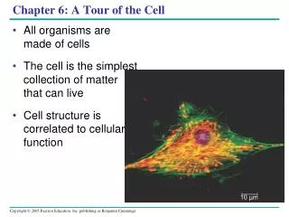
Learn how microscopes reveal the world of cells and discover the essential organelles that maintain cellular life. Understand the significance of cell size, shape, and structure within the context of cellular activities.

E N D
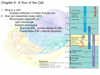
How We Study Cells Microscopes opened up the world of cells Characteristics of Microscopes • magnification: ability to make an image larger than actual size • resolution: power to show details clearly while enlarged (if poor, objects seem fuzzy) • constrast: accentuates different parts of the sample
Types of Microscopes • compound light - light passes through one or more lenses - object must be sliced thinly enough to be transparent - upper limitation is 2000X or 0.5 microns (um) in diameter - resolution limitation: 0.2 microns
II. Electron Microscopes (1950’s) - limited by physical characteristics of light - can magnify an image up to 200,000 X - beams of electrons produces enlarged image - resolution limitation: 0.002 nm across object
Types of Electron Microscopes • transmission electron microscope (TEM) - used to study internal structures - transmits a beam of electrons through very thinly sliced specimen stained with heavy metals - change density of cellular structures and electron transmission - dead specimens - 200,000 X magnification - black and white only Plant Cell
scanning electron microscope (SEM) - used to study surface structures - surface covered with thin film of gold - beam excites electrons on surface - produces three dimensional images - 100,000 X mag. - dead specimens only
Isolating organelles • Cell fractionation - take apart cells, separate major organelles • Ultracentrifuge - applies force 1 million times the force of gravity to separate further the cell organelles with the most dense at the bottom
CELL THEORY • all living things are composed of cells • cells are basic units of structure and function 3. all cells come from pre- existing cells
Activities of Life • Most everything you think of a whole organism needing to do, must be done at the cellular level… • reproduction • growth & development • energy utilization • response to the environment • homeostasis
Common features of all cells - plasma membrane (cell membrane) - cytosol: semi-fluid substance, holds cellular structures - cytoplasm: cystsol + organelles - cytoskeleton: microscopic protein fibers that keep cells shape - ribosomes: make proteins - DNA: controls all cell activities
Cell Size - most cells are 5-50 microns surface area ratio (limits size of cells) inside of cell grows faster: cubed (V = L x W x H) outside of cell grows slower: squared (A = L x W)
Surface Area Example • Cells must be small to maintain a large surface area to volume ratio • Large S.A. allows rates of chemical exchange between cell and environment Small Intestine: highly folded surface to increase absorption of nutrients • Villi: finger-like projections on small intestine wall • Microvilli: projections on each cell
Surface Area ExampleRoot hairs increase surface area for water and mineral absorption
Limits to Cell Size • Metabolic requirements set uper limit of size • In large cell, cannot move material in and out fast enough to support life
How to get Bigger • Become multi-cellular
- most spherical or cuboidal - different shapes reflect function dermal epidermal cells white blood cells goblet cell red blood cells nerve cell Cell Shape
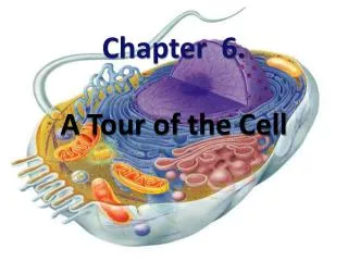
Eukaryotic CellsAnimal Plant Cytoplasmic channels- Connnect adjacent Cell’s cytoplasm
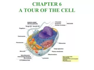
Cell Structure Main components of eukaryotic cells • cell membrane (outer boundary) • nucleus (control center) • cytoplasm (material between nucleus and membrane)
Nucleus - control center of cell: directs all cell activities - contains DNA - continuous with rough ER - site of DNA and RNA synthesis - located in center of most cells
Structure: - nuclear matrix - protein skeleton helps maintain nucleus shape - nuclear envelope (double membrane) - contains chromatin: combination of strands of DNA and protein - nuclear pores: control substance movement - nucleoplasm: dense, protein rich - nucleolus: region that forms ribosomal subunits
Cytosol(between membrane and nucleus) - Cytoplasm = cytosol + organelles • gel like material between • contains water, salts, organic molecules • in constant motion (cytoplasmic streaming) • holds organelles animation amoeba animation
Organelles Organelle: tiny structure that performs special functions in the cell to maintain life
Mitochondria • powerhouse of cell (cell respiration) • provides energy for cell in form of ATP • membrane bound • double membrane: • most numerous in cells which use a lot of energy (muscle) • self replicating, contain their own DNA - cristae: folds of inner membrane greatly enlarge surface area of inner membrane (more area for chemical reactions of respiration) - matrix: fluid filled inner compartment
Ribosomes • spherical structures which make proteins • not surrounded by membrane • composed of protein and rRNA • site of protein synthesis • free ribosomes: float in cytosol- make proteins used within cell • bound ribosomes: attached to rough ER- make proteins for export from cell (secretion)
Rough ER - ribosomes stuck to membrane surface - package proteins for secretion and inserted into ER - can be stored or exported to smooth ER - prominent in cells that make a lot of protein Smooth ER - no ribosomes - also stores and acts as an intercellular highway for proteins and enzymes involved in: - synthesis of steroids in gland - cell regulation of Ca levels in muscle - cells break down toxic substances in liver cells Endoplasmic reticulum: (ER)intercellular highwaycomplex membrane system of folded sacs and tunnels regulates protein traffic and performs metabolic functions
Golgi Apparatus • flattened system of membranes and sacs piles on each other (like pancakes) • very close to ER • processes, packages, and secretes proteins for transport (o other parts of cell (vesicles) and produce lysosomes • Cis face: receives vesicles • Trans face: ships vesicles animation
Steps of Protein Production and Transport • ribosomes make proteins on the rough ER- packaged into vesicles • vesicles transport the newly made proteins from the rough to the Golgi apparatus • in Golgi, proteins are processed and then packaged into NEW vesicles • vesicles move thru Golgi to cell membrane and release contents outside cell animation 2
Lysosomes • small round vesicles that contain digestive hydrolytic enzymes • formed from Golgi Apparatus • digest and remove waste from cell (old organelles, byproducts, bact., viruses) animation
Vacuoles • Storage of materials (food, water, minerals, pigments, poisons) • Membrane bound • Ex: food vacuoles, contractile vacuoles
Peroxisomes • contain different oxidative enzymes than lysosomes • break down toxic substances into H2O2 (remove H from substances and transfer them to O2) then converts H2O2 to H2O - detox alcohol and drugs - break down fatty acids • formed from proteins in cytosol, not Golgi • Glyoxysomes (fat tissues of plant seeds) FA sugar
Cytoskeleton (cell framework) • maintains shape and size of cell • composed of network of long protein strands located in cytosol • provides movement for organelles within cytosol • regulate biochemical activities
Cytoskeleton Structure • Intermediate Filaments (medium size fibers) - protein fibers coiled into cables - maintain shape of cell - permanent fixtures - anchor nucleus and organelles
Cytoskeleton Structure B. Microtubules (largest fibers) - long hollow coiled protein tubes (tubulin) - maintain shape and support cells - internal cell highways – move organelles thru cell - form centrioles, spindles (cell division) - motility (cilia and flagella)
flagella: long whip-like structures used for movement (motility) • cilia: short numerous hair like projections - movement - transport of substances across cell - signal receiving antenna for ex: ear drum: transmits sound waves respiratory tract: moves mucus etc. Motility requires interaction Basal body- anchors of cytoskeleton fibers with cilia/flagella to cell motor proteins.
Internal Organization 9 + 2 Arrangement dynein animation respiratory system animation
Centrosomes: region near nucleus • Microtubule organizing center • Contains centrioles • Used in cell division • Not found in plants or fungi • 9 + 0 arrangement
Cytoskeleton Structure C. Microfilaments (smallest fibers) - two strands fine protein (actin) intertwined - used in cytoplasmic streaming, muscle contraction, ameboid movement - smallest strands of cytoskeleton cytoplasmic streaming
Extracellular Matrix (ECM) • Outside plasma membrane • Composed of glycoproteins (collagen/ proteoglycans) • Strengthens tissues and transmits external signals to cell • Fibronectins/integrins: attach cells to ECM
Intercellular Junctions (Animal Cells) • Tight junctions: 2 cells are fused to form watertight seal • Desmosomes: “rivets” that fasten cells into strong sheets • Gap junctions: channels through which ions, sugar, small molecules can pass
Intercellular Junctions (Plant Cells) Plasmodesmata • Channels in plant cell walls that attach plasma nmembranes of bordering cells connect • Water, solutes, some proteins, RNA move through channels
Contain the same organelles as animal cells plus the following: 1. cell walls 2. vacuoles 3. plastids Plant Cells
Plant Cell wall • rigid covering of plant cells, algae, and some bacteria • composed of long chains of cellulose embedded in hardened lignin and pectin • very porous (O, H2O, CO2 easily pass through) • function: support & protection
- middle lamella laid first, formed from the cell plate during cytokenesis pectin: gluey substance holds cells together - primary cell wall forms next, expanded inside the middle lamella cellulose: structure and support - secondary wall constructed between the plant cell and primary wall after a maximum size has been reached and stops growing lignin: very stiff and hard, in woody plants in bark structure and support Structure
PlastidsConvert solar energy into chemical energy to be stored. 3 types (arise from proplastids) 1. chloroplasts- chlorophyll (green pigment) used in photosynthesis • chromoplasts- synthesize and store red, orange, and yellow pigments (give plants unusual colors) • leucoplasts- store starches, proteins, and lipids colorless