Download

1 / 25
260 likes | 347 Views
Explore the characteristics, historical impacts, transmission, and clinical manifestations of Yersinia pestis. Learn about the laboratory diagnosis, treatment, and prevention methods for this bacterium responsible for the deadly plague.

E N D
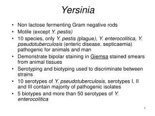
Yersinia • Y. pestis • Y. pseudoctuberculosis • Y. entercolitica
Species of Yersinia Bubonic Y pestis Plague Septicemic Pneumonic Food / water born diseases Y pseudotuberculosis 6 serotypes Y enterocolitica 27 serotypes
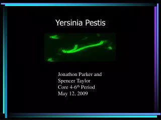
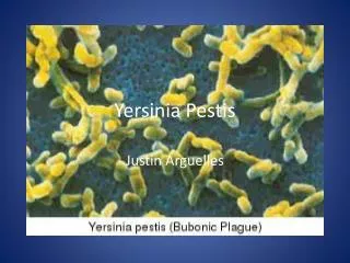
Yersinia pestis • Discovered by yersin and Kitasato • Short plump ovoid gram negative bacillus. • In Giemsa stained smears it shows bipolar staining (safety pin appearance). • It is capsulated, nonmotile.
Cultural characters • Aerobic and facultative anaerobic • Optimum temperature for growth is 27°C • Nutrient agar: transparent colonies • Blood agar: dark brown colonies due to absorption of hemin pigments
Ghee broth • Y.pestis when grown in a flask with oil or ghee floated on top, • a characteristic growth occurs which hangs down from under surface of ghee, resembling stalactites (stalactite growth)
Virulence factors • Fraction-I or F-I: Envelope protein which inhibits phagocytosis. • V and W proteins also inhibit phagocytosis. • Plague toxin: This is an endotoxin
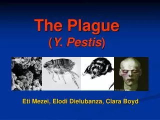
Historical Impacts • The first plague was in 542 B.C. and lasted almost 60 years. • The second and most severe pandemic was in the 14th century, also known as the Black Death. • The final and most recent pandemic occurred in 1894.
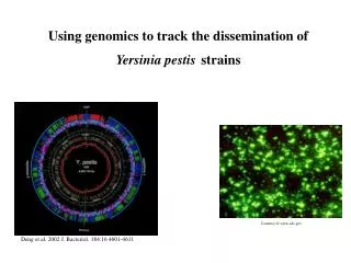
Transmission of the Plague • Plague is a zoonotic disease • Fleas found on rodents( xenopsiella cheopis) carry disease • Man to man transmission is through the droplets from coughing
How man acquire Plague ? Squirrel Rat Sylvatic plague Urban plague Flea Flea Infected flea bite human beings Spread via blood (Septicemic) Bubonic Plague Pneumonia & Meningitis Inhalation by other humans Pneumonic Plague
Diseases caused by Yersinia Pestis • Bubonic plague: based on the lymphatic system. • Septicemic plague: Centers in the bloodstream. • Pneumonic Plague: Centers in the lungs.
Plague: Clinical Manifestations Bubonic Pneumonic Septicemic Cervical bubo Gangrene Ecchymosis
Clinical feautres • Bubonic Plague • Constitutional symptoms • Lymphadenopathy • “bubo”: inguinal lymphnodes enlarged • May suppurate and drain • Septicemic Plague • Same terminal event of bubonic plague • Massive involvement of blood vessels leads to hemorrhages in skin and mucosa: BLACK DEATH
Primary pneumonic plague Organisms inhaled Lobular or lobar pneumonia Pulmonary necrosis Bacteremia Multiorgan seeding, failure Sepsis • Highly infectious form of plague
Laboratory diagnosis • Samples - blood, sputum, aspirated bubo fluid, splenic tissue on postmortem • Microscopy– Gram stain and Methylene blue stain: bipolar staining bacilli
Laboratory diagnosis • Culture – Blood agar, Ghee broth • Animal inoculation: Guinea pigs and rats injected subcutaneously with bubo fluid. Animals die within 2-5 days. • Antigen detection- F1 glycoprotein in bubo fluid and sputum • Serology –Antibodies to F1 glycoprotein appear at the end of 1st week of illness, and remain positive for several years • Rapid tests (PCR, DFA, etc) at reference labs
Identification features of Y pestis Ways of staining – bipolar staining Y pestis BA: Colonies are characteristically sticky CIN agar (cefsoludin irgasan- novobiocin) Gram stain
Treatment and Prevention • Tetracycline is drug of choice. • Vaccines: Two types • Killed vaccine: Haffkine’s vaccine. Virulent strain, Whole bacterial cell vaccine, 2000 million organisms/ml • Dosage: .05 ml S/C followed by 1ml after 7-14 days • Live vaccine: Avirulent strain of Y.pestis: Otten’s tjiwidej strain and Girard's 76 strain
Yersinia pseudotuberculosis & enterocolitica Facultative intracellular pathogens Pathogenesis Invade macrophages & epithelial cells Feces of wild and domestic animals Source of infection Ingestion of contaminated food or water Mode of transmission Y enterocolitica: pigs / dogs
Clinical manifestations Children < 5 yrs Acute enteritis with prolonged diarrhea Older children & adults Gastroenteritis Diarrhea Abdominal pain Terminal ileitis Mesenteric lymphadenitis LN enlarge Women 15-45 yrs Diarrhea with arthritis & erythema nodosum Liver, hemoglobinopathies, DM, Steroids Preexisting disease
Lab Diagnosis Y enterocolitica Specimens Stool samples Microscopy Gram negative coccobacilli – Gram stain Culture Non lactose fermenter – MAC agar Bulls eye colonies - CIN agar (cefsoludin – irgasan- novobiocin) Identification Biochemical tests
Tularemia • Francisella tularensis -Intracellular gram neg. coccobacillus. • It is also known as rabbit fever due to the fact that it may be transmitted to hunters and others who may have exposure to infected rabbits
Tularemia • Human infection may occur by handling or eating infected meat or drinking contaminated water Several forms: • Ulcers • Nodes • Pneumonic • usually ulceroglandular disease
Pasteurella multocida • Non motile, gram negative rod resembling yersinia. • Normal inhabitant og respiratory tract ofanimals like dogs cats cattle etc. • Human infections rare, but may cause wound sepsis following animal bites