Download
1 / 21
210 likes | 225 Views
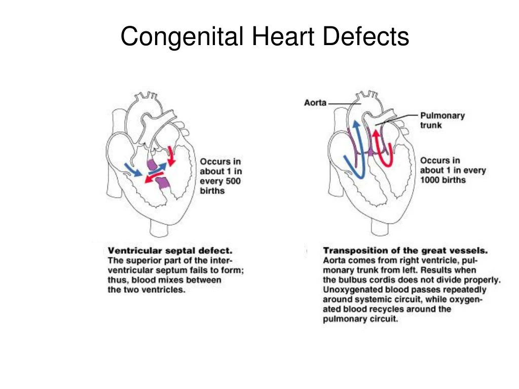
Explore congenital heart defects and the heart's electrical conduction system, including ECG tracing and components of blood. Learn about white and red blood cells, platelets, and the autonomic innervation of the heart.

E N D
Electrical Conduction System Sino Atrial (SA) Node – sup/post rt atrium Atrial Ventricular (AV) Node – inf rt atrium AV Bundle (of His) – sup IV septum L and R Bundle Branches – IV septum Purkinje Fibers – both ventricles
The Electrocardiogram Recording of the electrical activities in the heart P wave = Atrial Depolarization QRS complex = Ventricular Depolarization T wave = Ventricular Repolarization
Blood = Plasma + Formed (Cellular) Elements Plasma • ~ 55% blood volume • ~ 92% of plasma is water • High dissolved O2 content • Dissolved proteins • Minerals, glucose, ions. Cells • ~ 45% blood volume • RBCs (~ 99% of cells) • WBCs (~ 1% of cells)
The Proteins in Plasma • Albumins • 60% of plasma proteins. • Globulins • 35% of plasma proteins. • Fibrinogen • For clotting reaction, forms fibrin. • * serum = plasma without clotting proteins
Cellular Components RBCs (erythrocytes) ~ 99% of all cells. Hematocrit = % of blood occupied by cellular components. (packed RBC volume) Lacks nuclei, mitochondria and ribosomes. Anaerobic metabolism Life span = ~120 days
Scanning Electron Micrograph (SEM) of Erythrocytes or Red Blood Cells (RBCs) on the tip of a hypodermic needle.
White Blood Cells (Leukocytes) Granular Leukocytes • Neutrophils 70% of circulating leukocytes • Multi-lobed nucleus (3 or more), mobile phagocytes. • Eosinophils much less common • Bi-lobed nucleus with ‘orange’ staining granules. • Basophils relatively rare • Bi-lobed with dark staining granules, releases histamine.
Agranular Leukocytes • Lymphocytesprimary cell of lymphatic system • T-cells attack foreign cells directly. • B-cells produce antibodies. • Monocytes • Large nucleus, differentiate intoMacrophages.
n l e
Platelet cells (Thrombocytes) • Fragments of cells (Megakaryocytes) for clotting. Never Let Monkeys Eat Bananas
Arteries Arterioles Capillaries Heart! Venules Veins
Control of the Heart • Basic rate established by pacemaker cells inside the heart (myocardium) – called “intrinsic myogenic control” • Modified by Autonomic N.S. (ANS) • Para: decreases rate via the Vagus n. X. • Sym: increases heart rate and force of contraction via cardiac accelerator n.