Download
1 / 20
230 likes | 675 Views
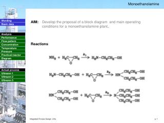
Hypersensivity Reactions. Allergies Greek = altered reactivity 1906 – von Pirquet coined term: hypersensitivity Hypersensitivity reactions – ‘over reaction’ of the immune system to harmless environmental antigens. Definition.

E N D
Hypersensivity Reactions Allergies Greek = altered reactivity 1906 – von Pirquet coined term: hypersensitivity Hypersensitivity reactions – ‘over reaction’ of the immune system to harmless environmental antigens
Definition • Hypersensitivity refers to undesirable (damaging, discomfort-producing and sometimes fatal) reactions produced by the normal immune system. • Hypersensitivity reactions require a pre-sensitized (immune) state of the host. • Hypersensitivity reactions can be divided into four types: type I, type II, type III and type IV, based on the mechanisms involved and time taken for the reaction.
Hypersensitivity reactions – originally divided into 2 categories: immediate and delayedIn 1968 Coombs & Gell defined the 4 types used today • Type I: classical immediate hypersensitivity • Type II: cytotoxic hypersensitivity • Type III: immune-complex mediated hypersensitivity • Type IV:cell mediated or delayed hypersensitivity
Hypersensitivity reaction depends on: 1) chemical nature of allergen 2) route involved in sensitization ie inhalation, ingestion, injection… 3) physiological state of individual / genetic potential
Type I (Dr Koshak will take it in details) • Common among population in developed nations • Prerequisite: need prior sensitization to antigen • the binding of antigen to antigen specific IgE bound on mast cells • Rapid liberation of active chemicals such as histamine and serotonin
Typical responses to these chemicals: • Increased capillary permeability Urticaria [hives] • Excessive mucus production Allergic rhinitis [hay fever] Diarrhea or vomiting Asthma
Type II • Type II Hypersensitivity • Type II hypersensitivity is also known as cytotoxic hypersensitivity and may affect a variety of organs and tissues. The antigens are normally endogenous, although exogenous chemicals (haptens) which can attach to cell membranes can also lead to type II hypersensitivity.
Type II • Drug-induced hemolytic anemia, granulocytopenia and thrombocytopenia are such examples. The reaction time is minutes to hours. Type II hypersensitivity is primarily mediated by antibodies of the IgM or IgG classes and complement (Figure 2). Phagocytes and K cells may also play a role (ADCC)
Type II • small molecules bound to cells and make a structure perceived as foreign by immune cells [ blood transfusion reactions. Erythroblastosis fetalis] • Allergens create a situation that induces cytolysis or cytotoxicity. • Antibodies involved are IgG & IgM • Complement is also involved
Complement • Blood proteins – initiate a series of enzymatic reactions leading to the ‘fixing’ of complement fragments to the pathogen’s surface – tagging it for destruction • Allergens trigger the classical complement pathway: antibody binds to specific antigen recruitment of inflammatory cells, opsonization facilitating phagocytosis
Type III Hypersensitivity • Type III hypersensitivity is also known as immune complex hypersensitivity. • The reaction may be general (e.g., serum sickness) or may involve individual organs including skin (e.g., systemic lupus erythematosus, Arthus reaction), kidneys (e.g., lupus nephritis), lungs (e.g., aspergillosis), blood vessels (e.g., polyarteritis), joints (e.g., rheumatoid arthritis) or other organs.
Type III • This reaction may be the pathogenic mechanism of diseases caused by many microorganisms.
The reaction may take 3 - 10 hours after exposure to the antigen (as in Arthus reaction). • It is mediated by soluble immune complexes. They are mostly of the IgG class, although IgM may also be involved.
Type III • The antigen may be exogenous (chronic bacterial, viral or parasitic infections), or endogenous (non-organ specific autoimmunity: e.g., systemic lupus erythematosus, SLE).
Type III • soluble protein complexes found in blood bound to IgG [ when non human proteins are given therapeutically – can be side effect] • Cause acute inflammatory reactions • Immune complexes can become deposited in walls of small blood vessels in alveoli anaphylaxis
Type IV Hypersensitivity • Type IV hypersensitivity is also known as cell mediated or delayed type hypersensitivity. • The classical example of this hypersensitivity is tuberculin (Montoux) reaction (figure 5) which peaks 48 hours after the injection of antigen (PPD or old tuberculin). • The lesion is characterized by induration and erythema.
Type IV • caused by products of antigen-specific effector T cells • T cells undergo blastogenesis and cellular division production of reactive cells • Usually takes 24 – 28 hours • No histamine or chemically related substances are released from cells
Type IV hypersensitivity is involved in the pathogenesis of many autoimmune and infectious diseases (tuberculosis, leprosy, blastomycosis, histoplasmosis, toxoplasmosis, leishmaniasis, etc.) • granulomas due to infections and foreign bodies