Download
1 / 57
590 likes | 654 Views
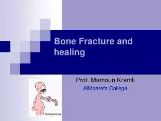
Understand bone anatomy, fracture patterns, healing mechanisms, and influence of implants. Learn about bone dynamics, strength, and healing cascade from inflammation to remodelling. Explore direct and indirect bone healing, fracture mechanics, and the impact of implants on bone biology.

E N D
Pathophysiology of fracture healing • Bone anatomy and biomechanics • Fracture patterns • Bone healing and blood supply • Influence of implants
Bone structure Four levels: • Chemical – molecular • Electron microscope – lamellae • Microscopic – Haversian systems • Macroscopic – compact and cancellous
Microscopy • Cortical bone • also “compact” and “lamellar” bone • Cancellous bone • spongy bone, woven bone.
Microscopy Haversian systems: • Lamellae interleaved with osteocytes in lacunae • Central canal with Blood vessel and lymphatics
Bone dynamics • Osteoblasts: mesenchymal, specialised adjacent to periosteum and endosteal areas • Osteoclasts: multinucleated giant cells, from bone marrow • Osteocytes: derived from osteoblasts, interlacunal connections, and entombed by their neighbours
Blood supply • Blood vessels- nutrient artery • Endosteal • Periosteal • Venous drainage
Bone Strength Compression Shear/tension
DESCRIBING THE FRACTURE • Mechanism of injury • Traumatic • Pathological • Stress • Pathological sieve
DESCRIBING THE FRACTURE • Anatomical site (bone and location in bone) • Configuration Displacement • three planes of angulation • translation • shortening • Articular involvement/epiphyseal injuries • fracture involving joint • dislocation • ligamentous avulsion • Soft tissue injury
MULTIFRAGMENTARY PROXIMAL- THIRD FEMORAL FRACTURE WITH SIGNIFICANT DISPLACEMENT OPEN? N/V INJURY?
Fracture mechanics • Spiral: Torsion Low energy
Fracture mechanics • Transverse: bending load
Fracture mechanics Oblique or transverse with butterfly: Compression + bend
Fracture mechanics Comminuted: High energy: combination • implosion • compression, • Bending • Torsion
Fracture healing Why do fractures unite? Because the bone is broken!
Healing cascade: indirect healing • Inflammation 0 – 5 days • Haematoma • Necrotic material • Phagocytosis • Repair: 5 – 42 days • Granulation tissue • Acid environment • Periosteum – osteogenic cells • Cortical osteoclasis • Remodelling • years
Cytokine release • Inflammatory mediators • Fibroblastic growth factor stimulates angiogenesis • TGF β initiates chondroblast/osteoblast migration • TGF β stimulates enchondral ossification
Healing cascade Late repair: • Fibrous tissue replaced by cartilage • Endochondral ossification • Periosteal healing » membranous ossification
Healing cascade Regeneration & remodelling • Replacement of callus (woven bone with lamellar bone) • Continued osteoclasis • Mechanical strain (Wolff 1892)
What is the difference between direct and indirect bone healing?
Indirect healing – healing by Callus • Unstable • Callus stabilises # • Direct healing between cortices
Robert Danis 1880 - 1962 • Plaque co-apteur, 1949 • Primary (direct) bone union “soudure autogène” • No callus
Direct bone healing – the response to rigid fixation • Temporary acceleration of Haversian remodelling • Only occurs in absolute stability of the fracture • Does not involve callus formation • Requires good blood supply
Direct bone healing Appositional healing • No gap • Osteons traverse # • Gap healing • Accurate apposition impossible • Vessels/mesenchymal • cells • Lamellar bone
Effect of implants on bone biology Absolute stability: Plates • Early reconstitution of macrocirculation • Plate footprint • Periosteal stripping • Titanium vv SS.
Effect of implants on bone biology Relative stability: IM nails • Reaming & blood supply • Periosteal reversal • Thermal necrosis
Effect of implants on bone biology Relative stability: External fixation • Pin configuration & rigidity of construct • Bone and thermal necrosis • infection
Cartilage and Bone • Cartilage--function, types, location • Bone Tissue--structure, types • Long Bone Structure and Development • Most common bone problems • Fractures • Osteoporosis
What is cartilage? • Skeletal tissue--maintains certain shape and form • Very resilient (bouncy or rubbery), mostly water • Grows fast--forms embryonic skeleton
Kinds of cartilage • Hyaline cartilage--most common, found in joints • Elastic cartilage--epiglottis, ear • Fibrocartilage--annular fibrosis of intervertebral disk, menisci of knee
M & M Figure 6.1
Bones provide: • Support and movement (limbs, axial skeleton) • Protection (skull bones) • Mineral storage • Blood cell development (long bone marrow) Bone is made up of: 35% collagen, ground substance and cells 65% calcium (hydroxyapetite)
Bone is alive!! Bone cell types: • Osteoblasts: Make and deposit components of bone extracellular matrix • Osteoclasts: Degrade and resorb bone for remodeling • Osteocytes: “watcher cells” Sit in bone and monitor its current status
Compact Bone Dense tissue at surface of bones Haversian canals Osteocytes in lacunae Highly vascularized Fig. 6.6, p. 138 Types of bony tissue
Trabecular (“spongy”) bone Trabeculae (oriented to give mechanical strength) Interior of long bones, skull bones Epiphyses of long bones Intramembranous ossification (osteoblasts lay down bone around blood vessels in connective tissues of dermis (after 8 weeks of development) Types of bony tissue
Structure of a long bone Fig. 6.3, p. 135 • Diaphysis (shaft) • Epiphysis • Proximal • Distal • Compact bone • Spongy bone • Periosteum • Medullary cavity • Articular/hyaline cartilage • Epyphyseal (growth) plates
Endochondral Ossification Fig. 6.9, p. 141 • Cartilage model • Bone collar forms in diaphysis (dense bone) • Cartilage chondrocytes in center of diaphysis die and cartilage disintegrates • Periosteal bud enters diaphysis • Makes spongy bone at ends of diaphysis (primary ossification center) • Epiphysis begins to ossify (secondary ossification center) • Hyaline cartilage remains only at • Epiphyseal surfaces (articular surfaces of joints) • Epiphyseal growth plates between diaphysis and epiphysis (primary and secondary ossification centers on either side)
Endochondral ossification centers—newly formed bone within cartilage shown is stained red
“Dig holes” with hydrochloric acid Degrades calcium Phagocytize collagen fibers and dead osteocytes Line tubes (Haversian canals) left by osteoclasts Lay down new bone in circular concentric lamellae Unique to warm-blooded animals--dinosaurs??? Osteoclasts Osteoblasts
Bone Fractures • Treatment is reduction • Closed--set in place by physical manipulation from outside body • Open--surgical placement of pins or screws • Healing • Hematoma • Fibrocartilaginous callus • Bony calllus • Remodeling by osteoclasts/osteoblasts • Types of Fractures